4 Joint AIC - SILS Conference

Europe/Rome
Doriano Lamba (CNR - Istituto di Cristallografia)
Description

Dear Colleagues,

It is with great pleasure that I invite you to the fourth Joint Conference of the Italian Crystallographic Association (AIC) and the Italian Synchrotron Radiation Society  (SILS), which will be held in Trieste from 12 to 15 September 2022.

The event returns to take place in Trieste nineteen years after the first one, held in July 2003.

Let us restart after the COVID-19 pandemic !

It will be a unique event, full of high-profile scientific content and for all a pleasant opportunity to meet, in presence, friends, colleagues and collaborators.

The Conference will be organized in 4 plenary lectures, thirteen micro-symposia with 26 keynote presentations, 39 oral communications and 39 flash talks and will host the Round Table "Horizon-Europe and the Future of Research Infrastructures: Next Generation of Scientific Instrumentation, Tools and Methods". Four E-poster sessions will also allow the presentation and discussion of scientific contributions.

Trieste, "suspended in time and history", a hub of relations and a crossroads in the circulation of knowledge, has gained international prestige over the years for the excellence of its scientific system, thanks to its University and more than 30 research and higher education centers in the area.

Trieste is the European city with the highest density of researchers (37.1 research workers per 1.000 workers) and hosted the Euro Science Open Forum in July 2020. It became part of UNESCO Global Network of Learning Cities in 2020.

The destiny of Trieste, which has in the bora wind one of its identity traits, has always been linked to its strategic geographical position, between the Mediterranean and Central Europe, crossroads of trade, people and cultures from the Middle Ages to the present day.

Trieste, declared free port in the Habsburg Empire, is the Italian city where the memory of the Empress Elizabeth of Austria, better known as Sissi, is more alive and tangible.

In Trieste, Central European, Mediterranean and Balkan traditions come together to create a cosmopolitan and international atmosphere.

On behalf of the AIC and SILS community, I look forward to welcoming you in Trieste.

Dr. Doriano Lamba

Chair of the Organizing Committee

Participants
  • Alberta Terzi
  • Alberto Cassetta
  • Alberto Lopez
  • Alberto Morgante
  • Alessandra Forni
  • Alessandro Genoni
  • Alessandro Grinzato
  • Alessandro Pesaresi
  • Alessandro Tofoni
  • Alessia Bacchi
  • Alma Cioci
  • Andrea Caporale
  • Andrea Ienco
  • Andrea Ilari
  • Andrea Martini
  • Andrea Pigozzo
  • Angela Altomare
  • Angelo Chiatto
  • Annalisa Guerri
  • Annie Heroux
  • Antonella Costanzo
  • Antonio Chaves-Sanjuan
  • Arnaud Grandeury
  • Artur Tuktamyshev
  • Beatrice De Bonis
  • Beatrice Vallone
  • Benedetta Chrappan Soldavini
  • Benny Danilo Belviso
  • Boby Joseph
  • Bruno Mascagna
  • Carlo Meneghini
  • Carmine Invigorito
  • Carmine Talarico
  • Cecilia Pozzi
  • Charlie Hitchman
  • Chiara Massera
  • Chieh-Hsuan Tsai
  • CINZIA GIANNINI
  • Clarissa Albertazzi
  • Corrado Cuocci
  • Cristian Biagioni
  • Daniela Mauro
  • Danilo Marchetti
  • Danny Stam
  • Davide Ceresoli
  • Davide Comboni
  • Davide Salusso
  • Demi Vattovaz
  • Denis Scaini
  • Dmitry Chernyshov
  • Domenica Marabello
  • Donato Belmonte
  • Doriano Lamba
  • Egilberto Lombardi
  • Elena Simone
  • Elisa Borfecchia
  • Eliza Kramarska
  • Eloise Mastrangelo
  • Emanuele Pelucchi
  • Emanuele Priola
  • Emiliano De Santis
  • Emiliano Principi
  • Fabia Gozzo
  • Fabio Fantini
  • Federico Boscherini
  • Federico Nolasco
  • Ferdinando Bosi
  • Flavia Squeglia
  • Francesca Menescardi
  • Francesca Paoletti
  • Francesca Rossi
  • Francesco Ardizzone
  • Francesco Bonì
  • Francesco Dallari
  • Francesco Princivalle
  • Francesco Stellato
  • Francesco Tavani
  • G. Diego Gatta
  • Gabriele Giuli
  • Gangadhar Das
  • Gennaro de Vita
  • GIANCARLO PANACCIONE
  • Gianluca Santoni
  • Giorgio Bais
  • Giorgio Paolucci
  • Giorgio Rossi
  • Giovanni Bulfaro
  • Giovanni Pierri
  • Giuliana Aquilanti
  • Giuliano Casati
  • Giuseppe Chita
  • Giuseppe Cruciani
  • Giusy Tassone
  • Guido Righini
  • Gustavo Santiso-Quinones
  • Heike E. Riel
  • Hendrik Wuensche
  • Hugo L. Monaco
  • Ida Freda
  • Ilaria Carlomagno
  • Iztok Arcon
  • Jana Kolar
  • Janis Timoshenko
  • Jasper Plaisier
  • Jing Cao
  • Leonardo Martinelli
  • Lisa Baratelli
  • Lorenzo D'Amico
  • Lorenzo Metilli
  • Luca Sironi
  • Lucia Allara
  • Lucia Carlucci
  • Lucia Robbia
  • Luciano Marchio'
  • Luisa Barba
  • Marco Mazzorana
  • Marco Merlini
  • Marco Milanesio
  • Marco Ranocchiari
  • Marco Taddei
  • Mariam Maisuradze
  • Marianna Marchini
  • Mariele Romano
  • Marina Mapelli
  • Marta Morana
  • Marta Stefania Semrau
  • Martina Lippi
  • Maryam Abdolrahimi
  • Mateja Pisačić
  • Mathilde Reinle-Schmitt
  • Matteo Busato
  • Matteo Daldosso
  • Matteo Melegari
  • Matteo Pancaldi
  • Mattia La Fortezza
  • Mattia Lopresti
  • Maurizio Polentarutti
  • Mauro Gemmi
  • Max Maletta
  • Michele Casappa
  • Michele Cianci
  • Michele Prencipe
  • Mickael Morin
  • Min Li
  • Mirko Gianesella
  • Neal Hickey
  • Nicola Campomenosi
  • Nicola Corriero
  • Nicola Demitri
  • Nodoka Hara
  • Oliviero Cannelli
  • Ornela De Giacomo
  • Paola D'Angelo
  • Paola Prete
  • Paola Storici
  • Paolo Lotti
  • Paolo P. Mazzeo
  • Paolo Scardi
  • Patrizia Rossi
  • Pietro Delre
  • Pietro Roversi
  • Pradip Kumar Mondal
  • Raghurama Prabhakara Hegde
  • Riccardo Signorato
  • Rita Berisio
  • Rita De Zorzi
  • Roberta Totani
  • Roberto Bergamaschini
  • Roberto Marotta
  • Romualdo Troisi
  • Rosaria Bruno
  • Sabrina Nazzareni
  • Sakura Pascarelli
  • Samet Ocak
  • Sandeep Kumar Chaluvadi
  • Serena Rocchio
  • Serena Tedesco
  • Sharon Shwartz
  • Silvano Geremia
  • Simon Parsons
  • Simona Fermani
  • Simona Galli
  • Simone Pollastri
  • Sirio Consani
  • Sonia Covaceuszach
  • Stefano Da Vela
  • Stefano Mangani
  • Stefano Morasso
  • Stevan Vrbaski
  • Thanalai Poonsiri
  • Theo Battista
  • Thomas R. Schneider
  • Tommaso Battiston
  • Valerio Bellucci
  • Valerio Cerantola
  • Wilson Crichton
  • Zeinab Ebrahimpour
Conference Secretariat
    • 12:00 13:45
      Registration Building N/-/- - Meeting Room (Parco San Giovanni)

      Building N/-/- - Meeting Room

      Parco San Giovanni

      200
    • 14:00 14:30
      Conference Open Cerimony -/-/- - Teatrino Basaglia (Parco San Giovanni)

      -/-/- - Teatrino Basaglia

      Parco San Giovanni

      200

      Roberto Di Lenarda - University of Trieste - Dean
      Angela Altomare - Institute of Crystallography CNR, Bari - President AIC - Conference Chair
      Cinzia Giannini - Institute of Crystallography CNR, Bari - President SILS - Conference Chair
      Federico Boscherini - University of Bologna - Chair of the Conference Scientific Committee
      Doriano Lamba - Institute of Crystallography CNR, Trieste - Chair of the Conference Organizing Committee

      Conveners: Prof. Francesco Princivalle (Università di Trieste - Dipartimento di Matematica e Geoscienze) , Silvano Geremia (University of Trieste)
    • 14:30 15:30
      Plenary: 1 -/-/- - Teatrino Basaglia (Parco San Giovanni)

      -/-/- - Teatrino Basaglia

      Parco San Giovanni

      200
      Convener: CINZIA GIANNINI (CNR - Istituto di Cristallografia)
      • 14:30
        Quantum imaging with X-rays 1h

        Quantum optics has been very fruitful and led to many seminal achievements in fostering quantum technologies in a variety of fields including imaging and spectroscopy [1]. However, the focus of quantum optics is on the optical range of the electromagnetic spectrum. The extension of concepts of quantum optics into the x-ray range can lead to remarkable applications with enhanced performances with respect to the present methodologies that are used in x-rays science and technology [2]. Recently, the properties of the strong time-energy correlations of photons produced by the process of spontaneous parametric down conversion (SPDC) have been extended to the x-ray regime to demonstrate the ability to improve the visibility and the SNR of an image with a small number of photons in an environment with a noise level that is higher than the signal by many orders of magnitude [3]. Another quantum imaging modality called ghost imaging has been reported as well [4]. More recently, the observation of the interaction of a single photon with a beam splitter has been observed [5] and extension of other quantum effects such as the Hong-Ou-Mandel effect to sub-attosecond sub-Angstrom optical path metrology with x-rays have been proposed [6]. Ghost imaging with SPDC of x-rays into visible radiation has been discussed for the high-resolution imaging [7]. Thereby, opening the possibility for future application of x-ray imaging and phase sensing applications with low radiation dose.
        In my talk I will review the recent progress toward quantum x-ray imaging and discuss the potential advantages of using quantum technologies for x-ray sensing.

        Figure 1. Example for x-ray quantum imaging: Reconstruction of the image of the triple slit object by (A) quantum enhanced x-ray detection. (B) Classical imaging of the same object for comparison. In each of the panels the horizontal axis represents the relative position of the object and the vertical axis represents the number of events that are detected.

        [1] S. Mukamel et al. “Roadmap on quantum light spectroscopy”, J. Phys. B: At. Mol. Opt. Phys. 53, 072002 (2020)
        [2] R. Röhlsberger, J. Evers, and S. Shwartz, Quantum and nonlinear optics with hard x-rays, in Synchrotron Light Sources and Free-Electron Lasers: Accelerator Physics, Instrumentation and Science Applications, edited by E. J. Jaeschke, S. Khan, J.R. Schneider, and J.B. Hastings (Springer International Publishing Cham 2020), pp. 1399–1431, https://doi.org/10.1007/978-3-030-23201- 6_32.
        [3] S. Sofer, E. Strizhevsky, A. Schori, K. Tamasaku, and S. Shwartz, “Quantum enhanced x-ray detection”, Physical Review X, 9, 031033 (2019).
        [4] A. Schori et al. “Ghost imaging with paired x-ray photons”, Physical Review A ,97 , 063804 (2018).
        [5] A. Schori and S. Shwartz "X-ray ghost imaging with a laboratory source”, Optics Express, 25,
        14822 (2017)
        [6] E. Strizhevsky, D. Borodin, A. Schori , S. Francoual , R. Röhlsberger , and S. Shwartz, "Efficient Interaction of Heralded X-Ray Photons with a Beam Splitter ” Physical Review Letters 127, 013603

        Speaker: Prof. Sharon Shwartz (Physics Department and Institute of Nanotechnology, Bar Ilan University, Ramat Gan)
    • 15:30 16:00
      Coffee Break 30m -/-/- - Anfiteatro - Roseto (Parco San Giovanni)

      -/-/- - Anfiteatro - Roseto

      Parco San Giovanni

      200
    • 16:00 18:20
      Session: AIC and SILS Awards -/-/- - Teatrino Basaglia (Parco San Giovanni)

      -/-/- - Teatrino Basaglia

      Parco San Giovanni

      200
      Conveners: Dr Angela Altomare (CNR - Istituto di Cristallografia) , Dr Cinzia Giannini (CNR - Istituto di Cristallografia)
      • 16:00
        AIC - "Mario Nardelli" Prize ex- aequo 5m

        Award Ceremony

      • 16:05
        Crystal Engineering for storage and release of molecular active ingredients 20m

        The ever-increasing attention on environmental problems and sustainability has highlighted several issues related to the use of conventional pesticides in the agricultural industry and the use of more effective food preservatives for the development of active packaging against food waste. In the search for natural antimicrobial and insecticide alternatives, essential oils and their active components have emerged as promising candidates, although they suffer from some drawbacks related to their physical properties. We exploited different crystal engineering approaches to stabilize the active components of the essential oils into the solid-state, either trapping them into flexible metal organic frameworks via crystalline sponge methods[1,2] or designing novel cocrystals with the final aim of controlling their release into the environment[3–5]. Wherever possible, and particularly for cocrystals, mechanochemical syntheses were preferred to wet reaction strategies. Providing “greener” and potentially less-expensive strategies than traditional solution methods, mechanochemistry was dubbed by the International Union for Pure and Applied Chemistry (IUPAC) as one of the 10 chemical innovations that will change our world. An excursus on different case studies will be provided with an hint into the mechanistic description of the mechanochemical synthesis via time-resolved in situ monitoring X-ray powder diffraction (TRIS-XRPD).[6,7]

        [1] D. Balestri, P. P. Mazzeo, R. Perrone, F. Fornari, F. Bianchi, M. Careri, A. Bacchi and P. Pelagatti, Angew. Chemie Int. Ed., 2021, 60, 10194–10202.
        [2] P. P. Mazzeo, D. Balestri, A. Bacchi and P. Pelagatti, CrystEngComm, 2021, 23, 7262–7269.
        [3] P. P. Mazzeo, C. Carraro, A. Monica, D. Capucci, P. Pelagatti, F. Bianchi, S. Agazzi, M. Careri, A. Raio, M. Carta, F. Menicucci, M. Belli, M. Michelozzi and A. Bacchi, ACS Sustain. Chem. Eng., 2019, 7, 17929–17940.
        [4] F. Bianchi, F. Fornari, N. Riboni, C. Spadini, C. S. Cabassi, M. Iannarelli, C. Carraro, P. P. Mazzeo, A. Bacchi, S. Orlandini, S. Furlanetto and M. Careri, Food Chem., 2021, 347, 129051.
        [5] F. Montisci, P. P. Mazzeo, C. Carraro, M. Prencipe, P. Pelagatti, F. Fornari, F. Bianchi, M. Careri and A. Bacchi, ACS Sustain. Chem. Eng., 2022 doi:10.1021/ACSSUSCHEMENG.2C01257.
        [6] G. I. Lampronti, A. A. L. Michalchuk, P. P. Mazzeo, A. M. Belenguer, J. K. M. Sanders, A. Bacchi and F. Emmerling, Nat. Commun., 2021, 12, 1–9.
        [7] P. P. M. Mazzeo, M. Prencipe, T. Feiler, F. Emmerling and A. Bacchi, Cryst. Growth Des, 2022, doi:10.1021/ACS.CGD.2C00262.

        Speaker: Prof. Paolo Mazzeo (University of Parma)
      • 16:25
        The quest for ordered porosity in zirconium phosphonates: blood, sweat and tears (and powder X-ray diffraction) 20m

        The chemistry of metal phosphonates has a long history, started in 1978 when the first examples of zirconium phosphonates were reported [1]. From the structural point of view, metal phosphonates often adopt dense layered or pillared-layered arrangements, with the organic moieties residing in the interlayer region, hardly accessible to potential guest species. In fact, most metal phosphonates are non-porous materials. When porosity is present, it is usually the result of a disordered assembly of the structure and lacks the specificity desirable for many applications, such as gas sorption/separation and catalysis [2].
        The advent of metal-organic frameworks (MOFs), a class of porous and crystalline organic-inorganic materials, has sparked huge research interest, thanks to their high surface areas and well-defined pore size distribution. Most MOFs are based on carboxylate or N-heterocyclic linkers, and phosphonate-based MOFs only represent a rather small fraction of the thousands of MOF structures reported over the last 20 years [2]. Yet, the exceptional stability and insolubility of metal phosphonates are attractive features for practical employment.
        This contribution is concerned with the challenging development of zirconium phosphonates possessing ordered porosity, starting from the design and synthesis of suitable polyphosphonic linkers to escape the ubiquitous dense layered motif, through the identification of the synthetic conditions to obtain sufficiently (micro)crystalline products, and ending with the ab initio determination of crystal structures from powder X-ray diffraction data [3].

        [1] G. Alberti, U. Costantino, S. Allulli, N. Tomassini, J. Inorg. Nucl. Chem. 1978, 40, 1113.
        [2] S.J.I. Shearan, N. Stock, F. Emmerling, J. Demel, P.A. Wright, K.D. Demadis, M. Vassaki, F. Costantino, R. Vivani, S. Sallard, I. Ruiz Salcedo, A. Cabeza, M. Taddei, Crystals 2019, 9, 270.
        [3] M. Taddei, F. Costantino, F. Marmottini, A. Comotti, P. Sozzani, R. Vivani, Chem. Commun., 2014, 50, 14831.

        Speaker: Dr Marco Taddei (Università degli Studi di Pisa)
      • 16:45
        Best PhD Thesis - "Fiorenzo Mazzi" Prizes 5m

        Award Ceremony

      • 16:50
        Metal-Organic Frameworks as advanced porous materials for gas separation and water remediation. 10m

        The removal of contaminants from air and aquatic systems must be a priority research topic to re-establish ecosystem balance and secure a more sustainable future. In this context metal–organic frameworks (MOFs) have revealed them as excellent platforms for the removal of harmful species from both air and water. MOFs show great potential for applications in the area of environmental sustainability because their channels can be pre- or post-synthetically fine-tuned, in terms of size, shape, and functionality, resulting in a fascinating controlled host−guest chemistry. In addition, unlike other porous materials, MOFs allow for the use of X-ray crystallography to observe what happens within their channels. Single crystal X-ray diffraction leads to structural resolution that allows to understand and rationalize the adsorption mechanisms that conduct to an efficient capture or separation of hazardous species.
        In this work, MOF-based technologies are reported unveiling successful developments achieved in the adsorptive removal of inorganic or organic contaminants. A new emerging class of porous materials, which are the mixed component metal-organic frameworks known as multivariate-MOFs (MTV-MOFs), have been also studied. In these MTV-MOFs heterogeneity and complexity are installed at the service of application performances. The modular nature of MTV-MOFs opens substantial possibilities in the field of water remediation, as it makes possible to tailor their porosity with two or more different and cooperative functional groups, capable of acting synergistically to capture contaminants of very different nature at once.
        In-situ studies on single crystal X-ray diffraction by synchrotron radiation revealed the nature of the interactions of gases with MOF active sites. These results were used then to prepare rubbery mixed matrix membranes (MMMs) with MOFs as filler and new MMMs for liquid or gas phase capture/separation were produced and tested.

        [1] M. Mon, R. Bruno, J. Ferrando-Soria, D. Armentano, E. Pardo. Journal of Materials Chemistry A, 2018, 6, 4912-4947.
        [2] R. Bruno, M. Mon, E.Tiburcio, M. Viciano-Chumillas, L. H. G. Kalinke, J. Ferrando-Soria, D.Armentano, E. Pardo. Journal of American Chemical Society, 2019, 141, 34, 13601-13609.
        [3] C. Negro, H. Martínez Pérez-Cejuela, E. Simó-Alfonso, J. Manuel Herrero-Martínez, R.Bruno, D. Armentano, J. Ferrando-Soria, E. Pardo ACS Appl. Mater. Interfaces 2021, 13, 24, 28424–28432.

        Speaker: Rosaria Bruno (Università della Calabria)
      • 17:00
        Applications of Cryo-Electron Microscopy in Structural Biology 10m

        For many years, cryo-electron microscopy (Cryo-EM) was used only to determine low-resolution density maps of large proteins structure. However, thanks to recent technological developments, the resolution achievable with Cryo-EM reaches the same values as those obtained with X-Ray crystallography. Moreover, Cryo-EM can study flexible and functionally active macromolecules, which are impossible to study using X-Ray crystallography.
        Below are presented the structures of four different protein complexes which, due to their flexible and heterogeneous nature, could not be solved without the aid of cryo-EM

        Campylobacter jejuni serine protease HtrACj is implicated in bacterial virulence, stress response, proliferation, migration, and cell fate. We took a cryo-EM approach to understand how its unique architecture is responsible for a remarkable structural and functional plasticity that allows cells to respond to misfolded or mislocalized polypeptides rapidly. The 3D reconstruction pictures HtrACj as a dodecamer built of four trimers. The contact between the trimers is loose, which explains the dodecameric assembly's flexibility and mobility. Such flexibility allows the opening of the dodecamer to expose the proteolytically active site of the protease.

        Tetanus neurotoxin (TeNT) is the bacterial protein toxin that causes tetanus. Although a vaccine exists for tetanus, it remains a health concern owing to the observations of decreasing anti-TeNT antibodies-load in human serum after the vaccination, implying the need to look for other therapeutics. Therefore, we explored the possibility of neutralizing TeNT using human-derived fabs. The structure of the TeNT-Fabs complex shows the relative movement of the toxin domains and the Fab bounding. Identifying new TeNT epitopes will help in antibody design to block the insurgents of tetanus.

        Photosystem II-LHCII is a protein complex of the thylakoid membrane involved in photosynthesis and is formed by one or more LHCII trimers that bind in a stable (S) or moderate (M) manner to the PSII dimeric core (C2). To study the PSII-LHCII light acclimation, we collected two different datasets of PSII-LHCII grown in low and high-light conditions, which led respectively to a map of the stacked (C2S2M2)2, (C2S2M)2, and (C2S2)2 conformation, confirming that M trimers decrease with the increasing of light exposure. Furthermore, all these maps show the presence of flexible connections between the two PSII-LHCII and a different rotational offset between the two PSII-LHCII of the stacked conformation around the membrane plane's normal vector, responsible for modifying their interaction and energetic connectivity.

        Potato virus X (PVX) is a flexible filamentous plant virus from the Alphaflexviridae family that infects herbaceous plants. While PVX has a detrimental impact on the global potato economy, PVX can be genetically engineered to be used as a scaffold for nanotechnology or nanomedicine applications. The 2.2 Å resolution map, obtained using helical reconstruction, provides near-atomic details of the interaction between its coat-protein and the genomic RNA, thereby opening the avenue to the design of antiviral compounds. Furthermore, the structure of the virus particle shows that the N-terminal region is flexible and exposed to the solvent, suitable for the transport and delivery of epitopes.

        Speaker: Alessandro Grinzato (European Synchrotron Radiation Facility Grenoble)
      • 17:10
        Crystal-chemistry of the secondary minerals of the thallium-rich pyrite ores from the Apuan Alps (Tuscany, Italy) 10m

        The mineralogy of secondary assemblages deriving from the alteration of pyrite ore deposits from the southern sector of the Apuan Alps (Tuscany, Italy) has been investigated. Twenty-five mineral species have been identified so far; among them, eight, showing interesting crystal-chemical features, were fully characterized using X-ray diffraction, chemical analyses, and spectroscopic techniques [1]. The structural description of some previously known minerals (epsomite, wilcoxite, coquimbite, khademite, alum-(K), and voltaite) allowed to improve our knowledge about H-bond systems in sulfates and to assess their role in hosting potentially toxic elements. Moreover, this study permitted the identification of two new mineral species, giacovazzoite, $K_5Fe^{3+}_3O(SO_4)_6(H_2O)_9·H_2O$, from the Monte Arsiccio mine, and bohuslavite, $Fe^{3+}_4(PO_4)_3(SO_4)(OH)(H_2O)_{10}·nH_2O$ (5 < n < 24), from the Buca della Vena mine [2, 3].
        Giacovazzoite (Figure 1) is the natural counterpart of the β-Maus’ Salt. It is monoclinic, space group P21/c, with a = 9.48, b = 18.44, c = 18.05 Å, β = 92.6°, V = 3153.6 Å$^3$. Its crystal structure is characterized by a heteropolyhedral $[Fe^{3+}_3O(SO_4)_6(H_2O)_3]^{5-}$ cluster and by an interstitial complex with composition $[K_5(H_2O)_7]^{5+}$. The heteropolyhedral cluster is known in other minerals (metavoltine, scordariite, carlsonite) as well as in some synthetic compounds.
        Bohuslavite (Figure 1) is triclinic pseudo-hexagonal, space group P-1, with unit-cell parameters a = 13.38, b = 13.34, c = 10.86 Å, α = 92.8, β = 91.0, γ = 119.9°, V = 1675.7 Å$^3$. This mineral can be considered a new kind of microporous compound, with heteropolyhedral layers with composition $[Fe^{3+}_4(PO_4)_3O(OH)(H_2O)_{10}]$, decorated on both sides by $SO_4$ groups. Additional $H_2O$ groups are hosted in channels running along c as well as in the interlayers.
        This work stressed the complexity of sulfates, improving the knowledge of their systematics.

        [1] C. Biagioni, D. Mauro, M. Pasero Minerals. 2020, 10, 1092.
        [2] C. Biagioni, L. Bindi, D. Mauro, M. Pasero Phys Chem Miner. 2020, 47, 7.
        [3] D. Mauro, C. Biagioni, E. Bonaccorsi, U. Hålenius, M. Pasero, H. Skogby, F. Zaccarini, J. Sejkora, J. Plášil, A.R. Kampf, J. Filip, P. Novotný, R. Škoda, T. Witzke Eur J Mineral, 2019, 31, 1033.

        Speaker: Daniela Mauro (Università degli Studi di Pisa)
      • 17:20
        Best BA/MS Thesis - "Fiorenzo Mazzi" Prizes 5m

        Certificate Delivery Ceremony

      • 17:25
        Crystal chemical study of arsenates and vanadates of the brackebuschite supergroup from Valletta mine, Canosio, Val Maira (CN) 5m

        The aim of this study is the crystal chemical description of arsenates and vanadates of the brackebuschite supergroup, belonging to Valletta mine, in Canosio municipality (CN), Piedmont, Italy, through structure and composition determination.
        The Valletta mine is type locality of three new species of the brackebuschite supergroup: canosioite $Ba_2Fe^{3+}(AsO_4)_2(OH)$, grandaite $Sr_2Al(AsO_4)_2(OH)$ and lombardoite $Ba_2Mn^{3+}(AsO_4)_2(OH)$. As-rich minerals probably form by precipitation from hydrothermal fluids in an oxidizing environment. They are concentrated in quartz or calcite-rhodochrosite veins, often associated with baryte, or along the fault planes. They are present in orange-brownish and submillimetric crystalline aggregates.
        Minerals of the brackebuschite supergroup have general formula $A_2M^{3+}(TO_4)_2(OH)$, in which:
        - A = Pb, Ba, Sr, Ca;
        - M = Al, Fe3+, Mn3+ (Zn, Cu if an hexavalent is present in the T site);
        - T = V, As, P (S, Si).
        The crystal structure can be described as composed of infinite chains of sharing edge octahedra [M3+O6] which extends along the [010] direction. The octahedra chains are connected to each other by two tetrahedra sites [T5+O4] or (OH)- groups or a water molecule.
        Minerals of the brackebuschite supergroup were characterized by:
        - transmission optical microscopy, for the description of thin sections and the measurements of the optical properties;
        - scanning electron microprobe (SEM) with EDS and WDS;
        - single crystal X-ray diffraction;
        - Raman spectroscopy.
        Following the analyses it was possible to identify a new lombardoite polytype and a new mineralogical phase of the brackebuschite supergroup; it was called aldomarinoite, in honour of Aldo Marino, researcher and collector, and discoverer of the Valletta mine. Aldomarinoite has chemical formula $Sr_2Mn^{3+}(AsO_4)_2(OH)$ and has recently been approved by IMA (International Mineralogical Association).
        The results of this study show that there is a relation between the Raman shifts and the chemical composition of the analysed minerals which would allow identifying different species in terms of peaks position variation in the Raman spectra. This was possible because of the understanding of structural variations obtained through structure refinements.

        Speaker: Lisa Baratelli
      • 17:30
        Thermal and compressional behavior of the natural borate kurnakovite, (MgB3O3(OH)5·5H2O) 5m

        Natural borates are minerals of relevant economic interest, as they represent the main source of boron (i.e. since 2014, in the Critical Raw Material list for the Europe’s economy). The peculiar properties of boron opened several routes of applications in a large variety of industrial sectors, including glass, ceramics, electronics, metallurgy, textile, cosmetics and chemistry [1]. Moreover, boron also finds applications as a fundamental constituent of neutron radiation-shielding materials, which are used in nuclear energy plants, as well as in facilities for scientific research or medical applications [2]. The high capability of borates to act as neutron-shielding materials is due to the isotope 10B, representing about 20% of natural boron, having a high cross section (~ 3840 barns) for thermal neutrons, leading to the following reactions:

        $^{10}B + n \rightarrow \alpha + ^{7}Li + \gamma$

        In this light, kurnakovite (MgB3O3(OH)5·5H2O, a = 8.308(7) b = 10.599(2) c = 6.442(3) α = 98.85(3) β = 109.09(6) γ = 105.57(4), space group = $P\bar{1}$) was selected as a promising natural candidate in neutron radiation-shielding materials, based on Portland or Sorel cements [3]. Moreover, the absence of Na prevents the occurrence of alkali-silica reactions, which can decrease the durability and the mechanical strength of Portland cements [4].
        In this study, the thermal and compressional behaviour of kurnakovite was investigated (up to 13.65(5) GPa and in a T range 100(2) – 393(2) K) by means in-situ single-crystal synchrotron X-ray diffraction experiments, performed at the XRD1 beamline at Elettra (Trieste, Italy) and at P2.02 beamline at PETRA-III (Hamburg, Germany). The bulk thermal expansion coefficient was calculated based on the non-ambient T data, yielding to the value of $\alpha_{\nu} = 5.18(1) \cdot 10^{-5} K^{-1}$. The crystal structure showed the first evidence of collapse at T > 393 K and completely amorphized at 423 K, posing questions on the potential application as matrix-aggregates in radiation-shielding concretes [4]. The refined bulk modulus of kurnakovite resulted to be $K_{V0} = 35(3) $GPa-1. A reconstructive phase transition was observed in the range 9.23(5) – 11.11(5) GPa [4]. The high-P polymorph, kurnakovite-II, is still triclinic with a unit cell volume three times larger with respect to kurnakovite [4]. As observed in other borates (e.g. ulexite and colemanite [5,6]), only a fraction of trigonal-planar B increases its coordination to tetrahedral by bonding with a H2O-oxygen atom [4].

        [1] D. Bernhardt, I.I. Reilly, Mineral Commodity Summaries 2019, US Geological Survey, Reston, USA, 2019.
        [2] M.A. Glinicki, A. Antolik, M. Gawlicki, Constr. Build. Mater. 164 (2018) 731–738.
        [3] E. Derun, A. Kipcak, J. Radioanal. Nucl. Chem. 2012, 292.
        [4] F. Pagliaro, P. Lotti, T. Battiston, D. Comboni, G.D. Gatta, F. Cámara, S. Milani, M. Merlini, K. Glazyrin, H.-P. Liermann, Constr. Build. Mater. 2021, 266, 121094.
        [5] D. Comboni, F. Pagliaro, G.D. Gatta, P. Lotti, T. Battiston, M. Merlini, M. Hanfland, Constr. Build. Mater. 2021, 291, 123188.
        [6] P. Lotti, G.D. Gatta, D. Comboni, G. Guastella, M. Merlini, A. Guastoni, H.P. Liermann, J. Am. Cer. Soc. 2017, 100.

        Speaker: Tommaso Battiston
      • 17:35
        Combined Experimental and Computational Studies on Molecular Recognition among D-Glucose Molecules 5m

        D–glucose ($C_6H_{12}O_6$) is a strategic chemical for the agri-food and pharmaceutical industries, with a market that is expected to grow in next years. Two anhydrous $P2_12_12_1$ crystal forms, differing in the anomeric ($\alpha / \beta$) configuration, plus one hydrated form, are known to date [1]. Due to the presence of a high number of –OH groups with almost null rotational barriers, many low-energy structures are predicted for both anomers in computational studies [2]. Thus, the reasons for the observed conformational monomorphism in glucose are unclear. In this work (Figure 1), an extensive crystallization screening was carried out, which led to the discovery of a new acetonitrile solvate of the $\beta$ anomer that was crystallized at room temperature. Through high resolution (0.46 Å) single crystal X-ray diffraction experiments at T = 90(1) K, the most common $\alpha$ form was characterized, demonstrating a possible anomeric disorder, never reported before. The experimental charge density study of the crystalline anomer $\alpha$ was obtained from the high-resolution dataset, quantifying the relative strength of hydrogen bonds in the crystal [3]. Finally, Molecular Dynamics simulations of the crystal, liquid and solution states were carried out with the MiCMoS [4] and GROMACS [5] suite of programs at different temperatures. These complemented the static, space-time average picture of standard crystallographic methods with the analysis of the equilibrium dynamics in condensed phase, allowing us to shed new light on the nature of the cohesive forces in D–glucose.

        Figure 1. (a)$\alpha$–D–glucose single crystals obtained from EtOH:pentane. (b) Same, from EtOH:hexane. (c) Crystal structure of the newly discovered $\beta$–D–glucose $CH_3CN$ solvate, with the space occupied by the solvent shown as brown cavities. (d) Negative charge density Laplacian ($–\nabla ^2 \rho$ ) in the region of one hydrogen bond. Solid lines highlight regions of charge concentration.

        [1] F.E. Young J. Phys. Chem. 1957, 61, 616-619.
        [2] T. Beyer, T. Lewis, S.L. Price CrystEngComm. 2001, 3, 178-212.
        [3] E. Espinosa, C. Lecomte, E. Molins Chem. Phys. Lett. 1999, 300, 745-748.
        [4] A. Gavezzotti, L. Lo Presti, S. Rizzato CrystEngComm. 2022, 24, 922.
        [5] M.J. Abraham, T. Murtola, R. Schulz, et al., SoftwareX. 2015, 1-2, 19-25.

        Speaker: Dr Luca Sironi (Università degli Studi di Milano)
      • 17:40
        SILS - Young Scientist Prize 5m

        Award Ceremony

      • 17:45
        The health seen through collagen network: from disease to regeneration 20m

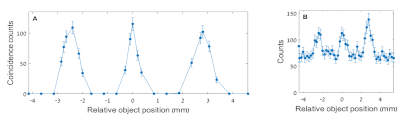
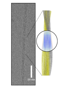
        In the last decades the extracellular matrix (ECM) has aroused growing interest thanks to its pivotal bio-physical interaction with cells, modelling the structure and function of tissues in relation to physiologic and pathologic stimuli. It is a complex fibrous network made by different components, but the main fibril-forming protein is type I collagen. Thanks to its tissue-specific morphology, its hierarchical structure and the functional domains, it supplies bio-physical support to cells attachment, tissue growth and re-modeling. From the molecular order, up to supramolecular scale, type I collagen is organized in triple helices assembled in fibrils and fibers, in accordance with a liquid crystalline arrangement at nanoscale, a quasi hexagonal packing observed in corneal tissue.[1] In order to study collagen arrangement and to deeply understand the role of its alteration at both atomic and nanoscale in pathology, we combined scanning small (SAXS) and wide angle (WAXS) X ray scattering microscopies, inspecting the structural features in the tissue over an extended area. For instance, mapping abdominal and popliteal aortic aneurysms biopsies by scanning X-ray microscopy led to the detection and co-localization of the nanometric structure of type I collagen, myofilament and elastin, organic components of the tissues. It were also identified pathological micro calcifications, whose crystalline phases have been identified.[2] Multiscale scanning microcopies were also employed for the structural characterization of intra- and inter-molecular features of type I collagen in diabetes minimal models for the study of the effect of glycation. In particular X-ray scattering (SAXS/WAXS) analyses conducted on decellularized bovine pericardia biotissues, soaked with different sugars (D-glucose, D-galactose, D-ribose) at increasing concentrations (0, 2.5, 5, 10, 20 and 40 mg/ml), and incubated at 37°C for 3, 14, 30 and 90 days, allowed to identify the sites of glycation and speed of glycation due to glucose/galactose and ribose.[3,4,5] Moreover the study of type I collagen by X-ray diffraction techniques have also demonstrated to be a useful tool in regenerative medicine. Indeed, its biocompatibility, activity and degradability, made collagen attractive as biomaterial for implantable medical devices. It was extracted from different collagen-rich tissues of distinct animal species by chemical and/or enzymatic processes and each biomaterial fabrication steps could modify the physiologic sub and supramolecular features of the protein, leading to different biomaterials. WAXS and SAXS techniques have revealed how manufacturing protocols deeply affect the structural characteristic of the biomaterial itself, and therefore its function, becoming fundamental tools to screen the suitable protocols, according to the tissue to regenerate. [6,7,8]
        [1] Sibillano, T., De Caro, L., Scattarella, F., Scarcelli, G., Siliqi, D., Altamura, D., Liebi, M., Ladisa, M., Bunk, O., & Giannini, C., Journal of applied crystallography, 2016, 49 Pt 4, 1231-1239 .
        [2] Giannini, C., Ladisa, M., Lutz-Bueno, V., Terzi, A., Ramella, M., Fusaro, L., Altamura, D., Siliqi, D., Sibillano, T., Diaz, A., Boccafoschi, F. & Bunk, O., IUCrJ, 2019, 6, 267-276.
        [3] Giannini C, Terzi A, Fusaro L, Sibillano T, Diaz A, Ramella M, Lutz-Bueno V, Boccafoschi F, Bunk O. J Biophotonics, 2019, 12(10)
        [4] Giannini, C., De Caro, L., Terzi, A., Fusaro, L., Altamura, D., Diaz, A., Lassandro, R., Boccafoschi, F. & Bunk, O., 2021, IUCrJ 8, 621-632
        [5] De Caro, L., Terzi, A., Fusaro, L., Altamura, D., Boccafoschi, F., Bunk, O. & Giannini, C., IUCrJ, 2021, 8, 1024-1034
        [6] A. Terzi, E. Storelli, S. Bettini, T. Sibillano, D. Altamura, L. Salvatore, M. Madaghiele, A. Romano, D. Siliqi, M. Ladisa, L. De Caro, A. Quattrini, L. Valli, A. Sannino, C., Giannini, Sci Rep., 2018, 8, 1, 1429
        [7] A. Terzi, N. Gallo, S. Bettini, T. Sibillan o, D. Altamura, L. Campa, M.L. Natali, L. Salvatore, M. Madaghiele, L. De Caro, L. Valli, A. Sannino, C. Giannini, Front Bioeng Biotechnol., 2019, 7, 203
        [8] D. Altamura, R. Lassandro, F.A. Vittoria, L. De Caro, D. Siliqi, M. Ladisa, C. Giannini, J.Appl. Cryst., 2012, 45, 869–87

        Speaker: Alberta Terzi (Institute of Crystallography-CNR)
      • 18:05
        SILS - PhD Thesis "Carlo Lamberti" Prize 5m

        Award Ceremony

      • 18:10
        A multi-technique hierarchical X-ray phase-based approach for the characterization and quantification of the effects of novel radiotherapies 10m

        Cancer is the first or second leading cause of premature deaths worldwide with an overall rapidly growing burden [1]. Standard cancer therapies include surgery, chemotherapy and radiotherapy (RT) and often a combination of the three is applied to improve the probability of tumour control. Follow up techniques, able to monitor and investigate the effects of therapies, are important for surveying the efficacy of conventionally applied treatments. They are also key for evaluating the curing capabilities and the eventual onset of acute or late adverse effects of new therapies. In this framework, this research project proposed the X-ray Phase Contrast Imaging - Computed Tomography (XPCI-CT) technique [2] to study and quantify the effects of novel RTs, namely Microbeam and Minibeam Radiation therapy (MRT [3] and MB [4]), and to compare them to the standard Broad Beam (BB) induced effects on brain and lungs in pre-clinical settings. MRT and MB deliver an array of highly collimated micrometric spatially fractionated X-ray beamlets issued from a synchrotron radiation source. To visualize with high sensitivity the effects of the treatment along and outside their path, a high-resolution and a full-organ imaging approach is necessary. XPCI-CT was here suggested and proven as a powerful imaging technique able to determine and quantify the effects of the radiation on normal and tumour-bearing tissues [5-6]. Moreover, it is shown as an effective technique to complement, with 3D information, the histology findings in the follow-up of the RT treatments. Using a multi-scale and multi-technique X-ray-based approach, we analysed the effects of RT delivery on healthy and glioblastoma multiforme-bearing rat brains as well as on healthy rat lungs. Ex-vivo XPCI-CT datasets acquired with isotropic voxel sizes down to 0.653 μm3 could distinguish, with high sensitivity, the idiopathic effects of MRT, MB and BB therapies. Histology, immunohistochemistry, synchrotron Small- and Wide-Angle X-ray Scattering and X-ray Fluorescence experiments were also carried out to accurately interpret and complement the XPCI-CT findings as well as to obtain a detailed structural and chemical characterization of the pathological and treatment-effect features. Overall, this multi-technique approach provided the recognition and differentiation of brain and lungs anatomical details down to the cellular level and identification of microscopic cancerous cell-clusters far from the main lesion. For brains, it was possible to discriminate tissue necrosis, tumour oedema, micrometric MRT-transections as well as high-density calcifications, identified, for the first time, as hydroxyapatite crystals with the coexistence of Fe deposits [5]. In lungs, radiation induced fibrosis and collagen noduli are visible. The former can be visualized as thickening of alveolar walls, expansion of alveolar spaces and destruction of their normal structures, which are replaced by irregular, abnormal air spaces and large areas of scarring [6]. The 3D nature of XPCI datasets was finally exploited to quantify the radio-induced pulmonary and brain lesions. This multi-technique approach appears to be well suited for investigating cancer development and radiotherapy effects on both the studied biological targets. In the future, other types of tumours and target organs will be considered and the method will be also tested for image-guidance during radiotherapy in preclinical research.

        [1] C.P. Wild, E. Weiderpass, B.W. Stewart, IARC 2020.
        [2] A. Bravin, P. Coan, P. and P. Suortti, Phys Med Biol 2013, 58, 1–35.
        [3] L.M. Smyth, S. Senthi, J.C. Crosbie, P.A. Rogers Int J Radiat Biol., 2016, 92, 6.
        [4] Y. Prezado, P. Deman, P. Varlet, G. et al. Rad Research, 2015, 184, 3.
        [5] M. Romano, A. Bravin, A. Mittone, et al. Cancers, 2021 19(13).
        [6] M. Romano, A. Bravin, M.D. Wright, et al. Int J Radiat Oncol Biol Phys 2022 112(3).

        Speaker: Dr Mariele Romano (Faculty of Physics, Ludwig-Maximilians-Universität, Garching, Germany)
    • 18:20 18:35
      Mineralogy 2022 -/-/- - Teatrino Basaglia (Parco San Giovanni)

      -/-/- - Teatrino Basaglia

      Parco San Giovanni

      200
      Convener: G. Diego Gatta (Univerdità degli Studi di Milano)
      • 18:20
        ‘Mineralogy 2022’: a worldwide celebration of mineralogy to highlight its importance in our everyday lives 15m -/-/- - Teatrino Basaglia

        -/-/- - Teatrino Basaglia

        Parco San Giovanni

        200

        Mineralogy 2022 (Min2022) belongs to the ‘International Year of Basic Sciences for Sustainable Development’ (IYBSSD2022) initiative approved by UNESCO. Major aims of Min2022 are i) to generate public interest in the science of matter and how it underpins most innovations and developments in our modern society, ii) attract young people to science through fascination with natural crystals, iii) illustrate the universality of science, iv) support the emergence of mineralogical societies in developing countries, especially those where natural resources are exploited, v) foster international collaboration between scientists worldwide, vi) promote education and research in mineralogy, crystallography and their links to other sciences, and vii) increase public awareness of the importance of natural resources.
        Mineralogy is a very active and rapidly evolving field with a tremendous impact on many facets our society. Besides being the basis of geology, mineralogy is closely related to crystallography, which applies the fundamental principles of crystal symmetry to mineral crystal structures. The mineral diversity and evolution are indicators of planetary evolution including the beginning of life on the Earth. Mineral occurrence is therefore a key factor in planetary sciences including the remote search for life in exoplanets. Mineralogy is also essential in searching for new sustainable resources (strategic metals, critical raw materials, etc.) either from natural deposits or human-made products including wastes. There is growing interest in understanding the interaction between the mineral world and the biosphere, which often involves major implications for human health. The search for solutions to the global climate change due to CO2 release in the atmosphere relies upon the promising approach for carbon capture and storage by minerals. Mineral-inspired new materials are designed to help with control of environmental pollution and recovery of treated resources.
        They main event will be a series of public lectures held during the General Meeting of the International Mineralogical Association (IMA) in Lyon during mid-July [1]. A dedicated website [2] and Twitter [3] are available to promote the events organized by the 39 national societies belonging to IMA, including the Società Italiana di Mineralogia e Petrologia (SIMP) [4] and its Gruppo Nazionale di Mineralogia (GNM) [5].

        [1] www.ima2022.fr
        [2] www.min2022.org
        [3] #Mineralogy2022
        [4] www.socminpet.it
        [5] www.socminpet.it/SIMP/GNM/

        Speaker: Prof. Giuseppe Cruciani (University of Ferrara)
    • 18:35 19:20
      Panel Discussion -/-/- - Teatrino Basaglia (Parco San Giovanni)

      -/-/- - Teatrino Basaglia

      Parco San Giovanni

      200

      Horizon-Europe and the Future of Research Infrastructures: Next Generation of Scientific Instrumentation, Tools

      Giorgio Rossi - University of Milano 18:35-18:45 NFFA-Europe Pilot: An Open Access Nanoscience Laboratory
      Giorgio Paolucci - Elettra-Sicrotrone Trieste 18:45-18:55 ELETTRA & Fermi Light Sources: status and upgrades
      Ornella De Giacomo - Central European Infrastructure Consortium - ERIC, Trieste 18:55-19:15 CERIC-ERIC: A Multidisciplinary Research Infrastructure
      Alberto Morgante - University of Triest 19:05-19:15 National Research Infrastructure Plan: CNR@Elettra2.0
      Questions Time 19:15-19:20

      Convener: Prof. Alberto Morgante (Physics Department,University of Trieste & CNR-IOM)
    • 19:20 20:00
      Meeting: Sezione AIC - Giovani Cristallografi DMGS Building O/-/- - Lecture Hall B (Parco San Giovanni)

      DMGS Building O/-/- - Lecture Hall B

      Parco San Giovanni

      200
    • 19:20 20:00
      Meeting: Sezione AIC - MacroMolecole Biologiche DMGS Building O/-/- - Lecture Hall A (Parco San Giovanni)

      DMGS Building O/-/- - Lecture Hall A

      Parco San Giovanni

      200
    • 20:00 21:30
      Welcome Party & Concert of Ricmanje Orchestra Wind Quintet 1h 30m -/-/- - Anfiteatro - Roseto (Parco San Giovanni)

      -/-/- - Anfiteatro - Roseto

      Parco San Giovanni

      200
    • 08:45 09:45
      Plenary: 2 DCPS Building C11/Ground Floor/- - Main lecture Hall (Università di Trieste)

      DCPS Building C11/Ground Floor/- - Main lecture Hall

      Università di Trieste

      184
      Convener: Prof. Simona Galli (Università degli Studi Dell'Insubria)
      • 08:45
        High Pressure Phase Transitions in Molecular Crystals 1h

        In the organic solid state, high pressure to 10 GPa leads to compression of intermolecular interactions and phase transitions. Pressure is therefore a very useful means for studying polymorphism in molecular solids. We have investigated hydrogen-bonded materials (e.g. amino acids) as well as very simple compounds (e.g. pyridine), and the talk will show how experimental structures of organic solids can be interpreted using semi-empirical and ab initio volume and energy calculations to develop an understanding of phase stability. Volume minimisation is the dominant driving force in almost all high-pressure phase transitions, although the relief of unfavourably compressed contacts can also play a role. Analysis of volume changes are therefore critical in the interpretation of phase transitions at high pressure, and we have developed a Monte Carlo algorithm for calculation of occupied (‘network’) and unoccupied (‘void’) space in crystal structures [1]. The variation of the volumes of the voids and the network of intermolecular contacts with pressure sensitively reveals discontinuities associated with first and second order phase transitions, providing insight into the effect of compression. The method is shown to be especially useful for the correlation of high-pressure crystallographic and spectroscopic data, illustrated for naphthalene, where a phase transition previously detected by vibrational spectroscopy, and debated in the literature for over 80 years, has been revealed unambiguously in crystallographic data for the first time.


        Figure 1. Variation of packing coefficient with pressure. Inset: loss of interstitial void space in naphthalene at high pressure.

        [1] C.J.G. Wilson, T. Cervenka, P.A. Wood, S. Parsons, Cryst. Growth Des., 2022, 22, 2328-2341.

        Speaker: Simon Parsons (University of Edinburgh)
    • 09:45 10:15
      Coffee Break 30m DCPS Bulding C11/III Floor/- - Atrium (Università di Trieste)

      DCPS Bulding C11/III Floor/- - Atrium

      Università di Trieste

      200
    • 10:15 12:15
      MS: 1 Modern Integrative Structural Biology DCPS Building C11/III Floor/- - Lecture Hall A1 (Università di Trieste)

      DCPS Building C11/III Floor/- - Lecture Hall A1

      Università di Trieste

      50
      Conveners: Prof. Stefano Mangani (Università di Siena) , Prof. Beatrice Vallone (Università degli studi "Sapienza" di Roma)
      • 10:15
        Molecular Crosstalk between Fate Determination and Orientation in Epithelial Cell Divisions 30m

        In multicelular organisms, oriented cell divisions are fundamental for morphogenesis and homeostasis [1]. Division orientation is orchestrated by the microtubule-based motor dynein, which sustains all mitotic spindle functions. During cell division, cortical force generators connect epithelial polarity sites with astral microtubules, allowing dynein movements to orient the mitotic spindle meanwhile astral microtubules depolymerize. Complexes of the LGN and NuMA proteins, that are fundamental components of force generators, are recruited to the cortex by Gαi-subunits of heterotrimeric G-proteins. They associate with dynein/dynactin and activate the motor activity pulling on astral microtubules. I will present the structure of NuMA:LGN hetero-hexamers, and discuss their role in promoting by multivalent interactions the assembly of active cortical dynein/dynactin motors required to oriented divisions in polarized cells. I will also describe the bipartite interaction interface between NuMA and the light intermediate chain (LIC) of eukaryotic dynein, supporting the notion that NuMA acts as a dynein-activating adaptor in the mitotic processes of spindle organization and positioning. Collectively this work elucidates the basis for the structural organization of essential spindle orientation motors.

        Key words: cell division, self-renewal, molecular motors, multivalent interactions

        [1] G. Lechler T. and Mapelli M. Nat. Rev. Mol. Cell Biol. 2021 doi: 10.1038/s41580-021-00384-4.
        [2] Renna C, Rizzelli F, Carminati M, Gaddoni C, Pirovano L, Cecatiello V, Pasqualato S, Mapelli M. Structure 2020 doi: 10.1016/j.str.2020.04.017 .
        [3] Pirovano L, Culurgioni S, Carminati M, Alfieri A, Monzani S, Cecatiello V, Gaddoni C, Rizzelli F, Foadi J, Pasqualato S, Mapelli M Nat. Comm. 2019 doi:10.1038/s41467-019-09999-w

        Speaker: Dr Marina Mapelli (Istituto Europeo di Oncologia)
      • 10:45
        Structural Characterization of a post-mortem Amyloid Fibril from a Cat Kidney 30m

        Amyloidosis are systemic diseases characterized by deposition of misfolded proteins into cross-β amyloids in multiple organs in humans and animals. During chronic inflammation, serum amyloid A protein (SAA) levels increases, which result in AA amyloidosis. There is a extreme disease prevalence in captive animals, e.g. 70% in captive cheetah and 57-73% in domestic short hair cats kept in shelters [1], and therefore a prion-like mechanism in amyloid formation has been proposed to explain the horizonal transmission od the disease.
        Herein, we present the 3.3 Å resolution cryo-EM structure of fibrils from renal tissue of a cat affected by severe AA amyloidosis in a shelter (Fig. 1) [2]. The fibril is assembled from two twisted proto-filaments, each comprising 76 residues. Amyloid fold and fibril assembly differ from previously reported human and mouse ex vivo AA amyloid structures. Almost identical fibril sequences and similar disease prevalence in related captive cheetah suggest that the structure reported here may depict the prion agent responsible for the high AA amyloidosis prevalence in these two related felids.

        Figure 1. Cryo-EM image of a single fibrilwith the reconstructed map and model

        [1] F. Ferri, S. Ferro, F. Porporato, C. Callegari, C. Guglielmetti, M. Mazza, M. Ferrero, C. Crinò, E. Gallo, M. Drigo, L.M. Coppola, G. Gerardi, T. Schulte, S. Ricagno, M. Vogel, F. Storni, M.F. Bachmann, A. Vogt, S. Caminito, G. Mazzini, F. Lavatelli, G. Palladini, G. Merlini, E. Zini. BioRxiv 2022.05.04.490646. 2022
        [2] T. Schulte, A. Chaves-Sanjuan, G. Mazzini, V. Speranzini, F. Lavatelli, F. Ferri, C. Palizzotto, M. Mazza, P. Milani, M. Nuvolone, A. Vogt, G. Palladini, G. Merlini, M. Bolognesi, S. Ferro, E. Zini, S. Ricagno. bioRxiv 2022.05.09.491126. 2022

        Speaker: Dr Antonio Chaves Sanjuan (Università degli Studi di Milano)
      • 11:15
        Insights on the structural determinants of proBDNF V66M variant, a modifier in neuropsychiatric disorders severity. 15m

        The human genetic variant BDNF (V66M) represents the first example of neurotrophin family member that has been linked to psychiatric disorders. In order to elucidate structural differences that account for the effects in cognitive function, this hproBDNF polymorph was expressed, refolded, purified and compared directly to the WT variant for the first time for differences in their 3D structures by DSF, limited proteolysis, FT-IR and SAXS measurements in solution. Our complementary studies revealed a deep impact of V66M polymorphism on hproBDNF conformations in solution. Although the mean conformation in solution appears to be more compact in the V66M variant, overall, we demonstrated a large increase in flexibility in solution upon V66M mutation. Thus, considering that plasticity in IDR is crucial for protein function, the observed alterations may be related to the functional alterations in hproBDNF binding to its receptors p75NTR, sortilin, HAP1 and SorCS2. These effects can provoke altered intracellular neuronal trafficking and/or affect proBDNF physiological functions, leading to many brain-associated diseases and conditions such as cognitive impairment and anxiety. The structural alterations highlighted in the present study may pave the way to the development of drug discovery strategies to provide greater therapeutic responses and of novel pharmacologic strategy in human populations with this common polymorphism, ultimately guiding personalized medicine for neuropsychiatric disorders [1].

        [1] S, Covaceuszach, L.J. Peche, P.V. Konarev, J. Grdadolnik, A. Cattaneo, D. Lamba. IJMS, under revision, 2022

        Speaker: Dr Sonia Covaceuszach (CNR - Istituto di Cristallografia)
      • 11:30
        C-terminus of the histone-lysine N-methyltransferase NSD3 characterized by small-angle X-ray scattering 15m

        Nuclear receptor binding SET domain (NSD) proteins, a family of three histon lysine methyl transferases, are considered decisive for suppressing currently lethal cancer diseases.[1] Aside from the well characterized catalytic SET domain, NSD have multiple potential chromatin-binding motifs that are clinically relevant, such as the proline-tryptophan-tryptophan-proline (PWWP), the plant homeodomain (PHD) and the adjacent Cys-His-rich domain (C5HCH) located at the C terminus. The crystal structure of the individual domains is available and has allowed initiating drug-designing of potential inhibitors, but the analysis of the intra-domain features and the characterization of mutual domain conformations has been hindered by the intrinsic flexibility of larger constructs. We have obtained the first structural characterization of the NSD3 C-terminal region comprising PWWP2, SET and PHD4 domains, by using solution small-angle X-ray scattering (SAXS). The challenging task of modelling flexible systems has been faced by complementing SAXS data on two multiple-domain NSD3 constructs with size-exclusion chromatography and advanced computational modelling. Structural models predicted by state-of-the-art homology modelling based on machine learning have been validated in direct space, by comparison with the SAXS-derived molecular envelope, and in reciprocal space, by reproducing the experimental SAXS profile. Selected models have been refined by molecular dynamics simulations, where the ab initio molecular envelope calculated from SAXS data represents an additional potential. The role of S-adenosyl methionine and histone H3 peptide ARTKQTARKSTGGKAPGGC in determining the geometrical features of the interdomain conformation has been also elucidated, finding a dramatic effect of the first ligand in shrinking the SET-PHD4 region. This study shows how SAXS data can be used in synergy with advanced computational modelling technique to achieve a detailed structural characterization that sheds light on how NSD3 domains are interconnected in the C-terminus.

        Figure 1. Structural model of the NSD3 C-terminal region comprising PWWP2, SET and PHD4 domains (center), obtained by modelling SAXS data on the constructs PWWP2-SET and SET-PHD4.

        [1] M. Morishita, E. di Luccio, Biochim Biophys Acta, 2011, 1816, 158.

        Speaker: Dr Benny Danilo Belviso (Institute of Crystallography)
      • 11:45
        Directional explosion. A hybrid simulation study 15m

        Single particle imaging (SPI) using X-ray pulses has become increasingly attainable with the advent of high-intensity free electron lasers. SPI overcomes the need of a crystalline sample, but the extreme intensity of the X-ray pulse causes severe radiation damage to the molecule, leading to coulombic explosion [1]. Diffraction patterns are thus obtained from separate exposures of identical molecules, each in their own random orientation. Hence, a single diffraction pattern reflects a single view of the particle. The unknown orientation of the particle renders its 3D reconstruction problematic, complicated and expensive – in terms of large sample consumption and beam-time required. If additional information about the orientation of the protein in the instant of X-ray exposure could be recorded, it would be beneficial for the algorithms that find the relation between the diffraction images.
        One way to control proteins orientation prior to the interaction with the X-ray pulses is to make them interact with external electric field [2]. Unfortunately, in some cases, the field can be destructive for the protein structures, as positively and negatively charged moieties will be pulled in opposite directions, potentially leading to unfolding and therefore to artefacts on the reconstructed images. It has also been proposed that by measuring the directions of the ejected ions from the Coulomb explosion of the protein, the orientation of the protein could be determined a posteriori [3]. In our previous simulation study, it has been shown how the sulphur atoms from a lysozyme protein tend to follow similar trajectories in 150 independent simulations. Since lysozyme is a rather small protein, and most of the sulphurs are closed to the surface of the protein, it is hard to draw any conclusions of how well defined the trajectories of atoms heavier than N, C and O from a larger protein would be. To test if the findings from the simulations agrees with what happens in an experiment, one needs to design an experiment to test this aspect. In such experiment it is necessary to know how the distance from the surface of the protein affects the direction of the ion path. In the present study we are addressing this specific question. By placing a sulphur atom at different distances from a water surfaces, and measure sulphur trajectories we aim at understanding how close to the surface of a biomolecule an atom can be to give reliable orientation information. To this end, we developed a hybrid simulation approach employing a combination of non-local thermodynamic equilibrium and extensive classical molecular dynamics simulations. Based on our findings, we conclude that explosion data can aid spatial orientation in SPI experiments and could substantially improve the capabilities of the SPI technique.

        [1] Chapman, Henry N., Carl Caleman, and Nicusor Timneanu. "Diffraction before destruction." Philosophical Transactions of the Royal Society B: Biological Sciences 369.1647 (2014): 20130313.
        [2] Sinelnikova, Anna, et al. "Protein orientation in time-dependent electric fields: orientation before destruction." Biophysical Journal 120.17 (2021): 3709-3717.
        [3] Östlin, Christofer, et al. "Reproducibility of single protein explosions induced by X-ray lasers." Physical Chemistry Chemical Physics 20.18 (2018): 12381-12389.

        Speaker: Emiliano De Santis (Uppsala University)
      • 12:00
        Structural insights into the hYAP-hTEAD4 protein-protein interaction: an emerging target in cancer treatment 5m

        The Hippo pathway is a signaling network, regulating cell growth, proliferation, and apoptosis, involved in tissue homeostasis and organ size control. A central role in this pathway is played by the Yes-Associated Protein (YAP), a DNA transcription co-activator without an intrinsic DNA binding domain. Upon activation of the Hippo pathway, a core kinase-cascade mediates the intracellular signaling, leading to phosphorylation and subsequent degradation of YAP. In its hypo-phosphorylated status, YAP moves inside the nucleus where it interacts with various DNA-binding partners. In mammalian cells, YAP primarily binds all four Transcriptional Enhancer Associate Domains (TEAD1-4) [1]. TEAD transcription factors can induce gene transcription only upon interaction with YAP [2]. This protein-protein interaction (PPI) is essential for expressing Hippo pathway-downstream genes, modulating cell proliferation and apoptosis [2]. All human TEADs (hTEADs) have an acylation binding site, physiologically occupied by palmitic/myristic acid; even so, the functional role of acylation is yet not fully understood [3]. Dysregulation of the Hippo pathway is associated with tumorigenesis, thus targeting the YAP:TEAD interaction is an emerging, attractive therapeutic strategy in the oncology field [4]. The development of new modulators of this PPI is challenging, indeed very few YAP:TEAD4 inhibitors have been reported so far [5]. A relevant problem is the poor structural information available on this complex, limited to the characterization of the C-terminal YAP-Binding Domain of hTEAD4 (hTEAD4-YBD) in complex with a 40mer peptide, a fragment of the TEAD-Binding Domain of human YAP (hYAP-TBD). Aiming to expand the current structural understanding on this PPI, we developed reliable protocols for co-expression and co-purification of hTEAD4-YBD in complex with hYAP-TBD (L-complex) and with two shorter fragments, including 70 and 90 residues (S and M complex, respectively). All three complexes were crystallized but, despite optimization, crystals showed only poor diffraction in preliminary X-ray crystallographic analyses. Meaningful improvements were achieved by the microseeding technique, allowing to obtain diffraction quality crystals for both the S and L complexes. The structure of the S complex was determined to 2.5 Å resolution, in the trigonal space group P3121 with unit cell parameters a,b=164.58 Å and c=258.61 Å, whereas the L complex was obtained to 3.1 Å resolution. The crystal ASU consists of 12 heterodimers, showing new structural insights on hYAP:hTEAD4 PPIs. Recently, we further expanded the study of this complex to the characterization of hTEAD4-YBD in complex with full length hYAP (XL complex). We set up a production protocol and preliminary structural characterization through bioSAXS, cryo-EM and X-ray crystallography are currently ongoing. Our results expand the current knowledge on the downstream effectors of the Hippo pathway, providing novel information to design hYAP:hTEAD4 PPI modulators.

        Speaker: Prof. Cecilia Pozzi (Dipartimento di Biotecnologie, Chimica e Farmacia, Università degli Studi di Siena)
      • 12:05
        Unravelling the regulation pathway of photosynthetic AB-GAPDH 5m

        Photosynthetic glyceraldehyde 3-phosphate dehydrogenase (GAPDH) is a key enzyme of the Calvin-Benson cycle. In higher plants different photosynthetic GAPDHs exist: the most abundant is formed by hetero-tetramers of A and B-subunits (AB-GAPDH). Being the major consumer of photosynthetic NADPH the enzyme is strictly regulated. The AB-GAPDH is indeed able to turn off its activity through oligomerization, a self assembly process mediated by the redox-sensitive B subunits tail called C-terminal extension (CTE). Typically, the fully inactive form is considered an hexadecamer A8B8, generated by the assembly of four A2B2-GAPDH tetramers [1].
        Our combined small angle x-ray scattering coupled with size exclusion chromatography (SEC-SAXS) and cryo-electron microscopy (cryo-EM) analysis revealed the presence of several AB-GAPDH oligomers [(A2B2)n-GAPDH oligomers with n=1, 2, 4 and 5] co-existing in a dynamic system and dependent on the solution conditions (activation/inactivation). We observed a great, and only partially explored, compositional and conformational heterogeneity that prevented us to obtain high resolution structures of AB-GAPDH oligomers (Fig. 1 A, B). The resolution we achieved was high enough to understand the inactivation/oligomerization mechanism common to all observed AB-GAPDH oligomers. The oligomerization was indeed obtained by the mutual exchange among adjacent B-subunits of their CTEs, which act as protruding hooks that dock into the active sites of adjacent subunits substantially blocking the access of the substrate.

        Speaker: Roberto Marotta (Istituto Italiano di Tecnologia)
      • 12:10
        Landscape of the ATP binding to neurotrophins: effects on conformation and dynamic modulation by divalent cations 5m

        Neurotrophic factors are involved in the maintenance and growth of neuronal populations. The first discovered member of neurotrophin (NT) family, Nerve Growth Factor (NGF), is essential for the development and maintenance of neurons and has a crucial role in activation of immune and endocrine systems and in the pain pathway. Mature NGF is expressed as precursor, proNGF, whose pro-peptide is an intrinsically unstructured domain (IUD). NGF and proNGF show different biological properties, the latter being involved in neuronal apoptosis. Additionally, the relative ratio of the two proteins is linked to neurodegeneration.
        The signaling pathway triggered by NGF involves the binding to TrkA and p75NTR receptors. Besides these receptors, proNGF also binds to sortilin receptor. Much is known about NGF’s role in neuronal physiology. However few reports have explored the role of essential endogenous ligands as modulators of NGF biological activity. Recent reports described the binding of ATP to NGF. The formed complex was proven protective versus death in neural cells. Various cellular studies investigated the ATP neurotrophic role in synergy with NGF and highlighted a crosstalk between NGF and ATP signaling systems.
        To fill the gap of understanding about the structural and mechanistic determinants of this binding, we used integrative structural biology to unveil for the first time the binding cartography of ATP to recombinant human NGF (rhNGF) [1]. Isothermal Titration Calorimetry (ITC) allowed us to measure a millimolar binding constant for ATP to rhNGF, and this was supported by 1H Saturation Transfer Difference NMR (1H STD-NMR) measurements. The 3D solution NMR structure of rhNGF was determined and exploited for a comprehensive binding study by ATP titration on rhNGF followed by 1H15N HSQC with related Chemical Shift Perturbation (CSP) analysis. The 15N NOESY collected at titration endpoint enabled us to identify new NOEs upon ATP binding. These were incorporated in MD simulations to identify the likely binding mode (position and orientation) of ATP on rhNGF. Surface Plasmon Resonance (SPR) allowed the effect of ATP on NGF binding to its receptors to be investigated.
        We also undertook a complementary biophysical study using Differential Scanning Fluorimetry (DSF), ITC, 1H STD-NMR and transferred NOESY NMR (trNOESY) measurements on the binding of ATP to recombinant human proNGF (rh-proNGF). Our results reveal a different binding profile for mature and precursor proteins. ITC suggested ATP binding to rh-proNGF with a micromolar dissociation constant. Integrative structural biology approach by Small Angle X-ray Scattering (SAXS), Hydrogen-Deuterium Exchange Mass Spectrometry (HDX-MS) and limited proteolysis showed that ATP binding induces a change in the conformation and/or dynamics of rh-proNGF, predominantly in the IUD pro-peptide. Interestingly, we have uncovered that the strength of ATP binding to rh-proNGF is modulated by Mg2+ and by their relative stoichiometric ratios.
        Combined, these results suggest a functional role for ATP in modulating the biological role of proNGF/NGF in health and disease states.

        [1] F. Paoletti, F. Merzel, A. Cassetta, I. Ogris, S. Covaceuszach, J. Grdadolnik, D. Lamba, S. Golič Grdadolnik 1., Comput. Struct. Biotechnol. J., 2021, 19, 2938.

        Speaker: Dr Francesca Paoletti (National Institute of Chemistry, Ljubljana, Slovenia)
    • 10:15 12:15
      MS: 3 Italy@EuXFEL DCPS Building C11/III Floor/- - Lecture Hall A3 (Università di Trieste)

      DCPS Building C11/III Floor/- - Lecture Hall A3

      Università di Trieste

      46
      Convener: Dr Sakura Pascarelli (EuXFEL Hamburg)
      • 10:15
        Megahertz-rate Ultrafast X-ray Scattering and Holographic Imaging at the European XFEL 30m

        During 2019, the novel DSSC 2D imaging detector [1] was commissioned at the Spectroscopy and Coherent Scattering (SCS) instrument of the European X-ray Free-Electron Laser (EuXFEL) [2]. To fully exploit the high repetition rate provided at EuXFEL (up to 2.25 MHz), the DSSC detector was designed to be the fastest one-megapixel camera available worldwide, providing single-photon sensitivity in the soft X-ray regime. As a test-bed for operation at megahertz rates, time-resolved magnetic X-ray scattering and holographic imaging experiments in solid state samples were chosen as representative [3]. Indeed, these two types of measurements allow to assess fundamental features of a 2D detector for X-ray science. For example, a high dynamic range is needed for recording the tiny variations in magnetic scattering induced by a laser pump, and holography requires measurements characterized by a high signal-to-noise ratio in order to provide well-defined image reconstructions. During this talk, we will present the obtained results, which were validated by measurements performed at other facilities (BESSY II, SOLEIL, MBI).
        As potential future application for high-repetition rate scattering and imaging experiments at free-electron lasers, we propose the use of light beams possessing orbital angular momentum (OAM), which are rapidly becoming a way for probing condensed-matter systems, even in the X-ray regime [4]. The wavefronts of such beams are characterized by an azimuthal angular dependence of the electric field phase, associated with an OAM topological charge $l \ne 0$.
        In this framework, OAM beams allow for novel kinds of dichroism experiments, paving the way for new spectroscopic tools in the fields of orbital physics and magnetism [5]. In particular, it has been demonstrated that, after the scattering of an OAM beam from magnetic structures featuring a non-uniform magnetization (like magnetic vortices), the far field intensity profile encodes the vortex symmetries in a way that depends on the sign and value of $l$, giving rise to magnetic helicoidal dichroism [6].
        On the other hand, for imaging purposes, the use of OAM beams can help in overcoming the Rayleigh criterion limit, so enhancing the theoretical resolution with respect to gaussian illumination ($l = 0 $) [7]. This feature has been recently tested at the DiProI beamline of the FERMI free-electron laser [8] by performing ptychographic experiments with a standard sample. As predicted, the ptychographic reconstructions with OAM beams showed a higher image resolution, and the retrieved illumination functions proved to be very sensitive to optical aberrations, providing the basis for new characterization and diagnostic tools.

        • on behalf of the collaboration supporting the beamtime “X-ray holography of ultrafast magnetism: femtosecond movies at the nanoscale” (EuXFEL proposal ID 2222)

        [1] M. Porro et al., IEEE Trans. Nucl. Sci. 2021, 68, 1334.
        [2] W. Decking et al., Nat. Photonics 2020, 14, 391.
        [3] N. Zhou Hagström et al., arXiv:2201.06350 [cond-mat.mes-hall].
        [4] Y. Shen et al., Light Sci. Appl. 2019, 8, 90.
        [5] M. Van Veenendaal and I. McNulty, Phys. Rev. Lett. 2007, 98, 157401.
        [6] M. Fanciulli et al., Phys. Rev. Lett. 2022, 128, 077401.
        [7] F. Tamburini et al., Phys. Rev. Lett. 2006, 97, 163903.
        [8] E. Allaria et al., Nat. Photonics 2012, 6, 699.

        Speaker: Dr Matteo Pancaldi (Elettra Sincrotrone Trieste)
      • 10:45
        Ultra-fast structural dynamics in materials at extreme conditions 30m

        Planetary science is witnessing a revolution with the discovery of hundreds of extra-solar planets orbiting nearby stars [1]. Characterising such astrophysical objects requires the knowledge of physical properties of their main constituents at multi-Mbar pressures and few-eV temperatures (1 eV = 10 605 K). The precondition is the establishment of phase diagrams of these materials, in order to identify and characterize structural changes, phase transitions, metallization, and dissociation processes. Physical and chemical changes induced by the application of high pressures and temperatures can occur in a wide range of different time scales, thus capturing their nature and underlying mechanisms requires the use of adapted tools, capable to catch happening phenomena in static and dynamic regimes. Also, it was shown that materials structural response to pressure increase might be different under static or dynamic compression loads i.e. [2].
        Since the opening in 2019, HED Instrument at the European XFEL has persistently upgraded and today offers standard platforms for static and dynamic compression studies, at timescales spreading between minutes and seconds to micro- or nanoseconds. X-ray diffraction and emission spectroscopy are routinely coupled to diamond anvil cells (static and dynamic, dDAC) in a different range of sample environments. Relevant studies comprehend the synthesis of metastable iron hydrides, phase diagram of water, dynamic compression of high-Z compressible gases like Krypton, the spin states and electronic transitions in iron sulfates, carbonates, and melts and many more. Shock-compression will be also available by the end of 2022 and will be coupled initially to x-ray diffraction and large area detectors. The presentation will focus on our most recent and interesting results. An outlook towards the future will elucidate ongoing projects and expected outcome.

        [1] Mayor, M. and Queloz, D. A Jupiter-mass companion to a solar-type star. Nature 1995, 378, 355–9.
        [2] Pépin, C. M. et al. Kinetics and structural changes in dynamically compressed bismuth. Phys. Rev. B 2019, 100, 060101

        Speaker: Dr Valerio Cerantola (EuXFEL Hamburg)
      • 11:15
        First high energy and temporal resolution pump-probe RIXS at the EuXFEL 15m

        High-resolution Resonant Inelastic X-ray scattering (RIXS) and ultrafast pump-probe techniques using optical or THz radiation have nowadays reached a prominent role for the investigation of strongly correlated materials.
        At the intersection of the two worlds, the development of a high energy and high temporal resolution RIXS instrument at the SCS beamline of the European XFEL is a cornerstone in the field of x-ray techniques. After the successful commissioning of the User Consortium Heisenberg RIXS (hRIXS) spectrometer in May 2021, we have recently carried out first pump-probe (pp) RIXS experiments. As a test case, we selected the prototypical cuprate parent compound La$_2$CuO$_4$ (LCO) and NiO, pumped with an optical laser above the Mott gap. The pp-RIXS measurements at the Cu L$_3$ and Ni L$_3$ edges were performed with a 113 kHz pulse repetition rate, reaching a temporal resolution of ≈100 fs and an energy resolution of ≈ 93 meV and ≈ 80 meV for the Cu and Ni L3 edges, respectively. Exploiting full control over laser power and delay between optical and x-ray pulses, we acquired spectra changing the pump delay between 0.1 ps and 5 ps, and the laser fluence on the sample from 1 mJ/cm$^2$ up to 35 mJ/cm$^2$.
        We observed strong changes in the orbital and charge-transfer excitations driven by the optical pumping in both samples, clearly scaling with the laser fluence and with a sub-ps dynamics. The results of this first high-resolution ppRIXS commissioning represent a major success and opens the route to ppRIXS studies on strongly correlated materials at the European XFEL.

        Speaker: Leonardo Martinelli (Dipartimento di Fisica, Politecnico di Milano)
      • 11:30
        Fast 4D X-ray Imaging at European XFEL 15m

        Fast 4D X-ray imaging is an emerging method that allows seeing fast-changing phenomena in opaque samples. This novel method will enable capturing stochastic phenomena happening at km/s speed into 3D volumetric videos. 2D X-ray phase-contrast microscopy with a sampling rate of 1.128 MHz has already been demonstrated at the European XFEL [1] by attaining videos with micrometer-scale spatial resolution. The contrast and spatial resolution are superior to previous synchrotron MHz microscopy due to the high brilliance of the source. 4D X-ray imaging at MHz rate is attained by dividing the main beam into many beamlets, each having a different view angle of the sample [2]. Therefore, this method requires a main beam with an even higher brilliance, and it is enabled by the new generation of XFEL sources. MHz 4D X-ray imaging has never been possible before and for this reason, there are fast phenomena never captured in 3D. These include modern emerging technologies, such as additive manufacturing, bioprinting, and new material production, whose development will benefit from fast volumetric imaging by probing the high-speed dynamics happening in such systems. MHz 4D X-ray imaging will also improve the understanding of industry and society relevant phenomena such as the formation and propagation of cracks in metals and aerospace alloys, which modeling is currently left to simulations and speculations with no possibility for direct observation. Experimental observation would facilitate the development of branches of science depending on fast phenomena such as material science, with the specific example of advanced and high-performance alloys.

        [1] P. Vagovič et al.," Optica. 2019, 6, 1106-1109.
        [2] P. Vagovic, V. Bellucci, P. Villanueva-Perez, W. Yashiro, Patent pending, Application number: EP21200564.9

        Speaker: Valerio Bellucci (European XFEL)
      • 11:45
        Microsecond dynamics in complex liquids with MHz XPCS 15m

        Complex liquids are a broad family of materials that cover key roles in several aspects of everyday life, from biological processes that can take place only in such environments to industrial applications that sees complex liquids either as a final product or as a fundamental manufacturing step. From a physicist perspective most of the more interesting phenomena take place at inter-particle distances, which for proteins and nanoparticles correspond to few nanometers, accessible only via X-ray based techniques. Moreover, such processes are often connected to diffusion mechanisms, which for water-based systems implies timescales of the order of few microseconds. These time and spatial constraints pose a real challenge to current 3rd generation synchrotron sources, limiting experiments only to a handful of complex experiments on prototypical systems [1].
        The MHz repetition rate of the European XFEL matches perfectly with these timescales making it an ideal choice for this kind of experiments. Here we report the results from MHz X-ray Photon Correlation Spectroscopy (XPCS) experiments performed at the instruments MID and SPB/SFX [2,3], showing how it is possible to execute measurements both on prototypical charge-stabilized silica in water and on radiation-sensitive core (silica) – shell (PNIPAm) nanoparticles. Moreover, we can show that tuning the pulse intensity and repetition rate it is possible to control the radiation-induced heating of the system without necessarily damaging it, which, combined with the capabilities of the XPCS techniques, opens the way to the study of out of equilibrium dynamics.

        [1] Q. Zhang, E. M. Dufresne, S. Narayanan, P. Maj, A. Koziol, R. Szczygiel, P. Grybos,
        M. Sutton, and A. R. Sandy, Journal of Synchrotron Radiation, 2018, 25(5), 1408–1416
        [2] F. Lehmkühler, F. Dallari., A. Jain, M. Sikorski, J. Möller, A. Madsen, A. P. Mancuso, and G. Grübel et al. PNAS, 2020, 117, 39, 24110-24116.
        [3] F. Dallari A. Jain, M. Sikorski, J. Möller, A. Madsen, A. P. Mancuso, G. Grübel, Felix Lehmkühler et al. IUCrJ, 2021, 8, 5, 775-783,

        Speaker: Dr Francesco Dallari
      • 12:00
        Mapping excitation-specific electron, spin and nuclear dynamics in spinel Co3O4 thin films with femtosecond X-ray emission spectroscopy and diffraction 15m

        We present a combined femtosecond X-ray emission spectroscopy (fs-XES) and X-ray diffraction (fs-XRD) study of spinel Co3O4, a system representing a prototypical case of the intrinsic complexity of transition metal oxides, due to a correlated response of its nuclear, spin and electronic degrees of freedom.
        This material has a normal spinel structure where Co2+ and Co3+ centres are surrounded by O2- anions and respectively occupy tetrahedral and octahedral sites with a 1:2 stoichiometric ratio. As a consequence, the electronic configuration of the metal centres is characterized by paired and unpaired d-electrons, and it determines the presence of Mott-Hubbard and charge transfer gaps close in energy in the Visible-NIR range, making it promising candidate for photovoltaic and photocatalytic applications [1].
        A full understanding of the material’s photodynamics and the excitation wavelength influence on its photoresponse calls for investigation techniques able to follow the charge carrier relaxation with ultrafast time resolution and with electronic and nuclear sensitivity. Here we excite the ligand-to-metal charge transfer (LMCT, 400 nm) and metal-to-metal charge transfer (MMCT, 800 nm) transitions of 27 nm Co3O4 thin films simultaneously mapping the lattice response with fs-XRD, and the spin and electronic transient configurations of the Cobalt centres with fs-XES. The experiment was performed at the FXE instrument of the European XFEL facility probing the system in grazing incidence with a pink beam centred at 9.3 keV and detecting the XRD signal with the LPD detector and the nonresonant Co Kα1,2 and Kβ1,3 emission lines using a Von Hamos spectrometer and Jungfrau detectors.
        Our joint investigation shows distinct decay pathways for the two excitation channels that are unaccessible with transient optical techniques. The results outline a radically different picture compared to previous time-resolved studies of Co3O4 [2], excluding a stepwise cascade mechanism in the charge carrier relaxation of the material and highlighting the relevance of electron-phonon coupling in the photocarrier response on ultrafast time scales [3].

        Speaker: Dr Oliviero Cannelli (EPFL)
    • 10:15 12:15
      MS: 7 Materials at Extreme Conditions: X-ray Crystallography and Beyond DCPS Building C11/III Floor/- - Lecture Hall A2 (Università di Trieste)

      DCPS Building C11/III Floor/- - Lecture Hall A2

      Università di Trieste

      50
      Conveners: Dr Giuliana Aquilanti (Elettra Sincrotrone Trieste) , Dr Davide Comboni (ESRF Grenoble)
      • 10:15
        Exploring Phase Diagrams, Structures and Properties with the Large Volume Press at the ESRF 30m

        The ESRF’s large-volume press beamline, ID06LVP, has been in User operation for 10 years (e.g. Guignard & Crichton, 2015). It complements the ESRF’s already wide-ranging extreme conditions portfolio by offering, primarily, in situ angle-dispersive diffraction-based experimentation with a multi-anvil device that operates in one-and two-stage modes. Through use of independent anvils; both normal compression, with high degrees of triaxiality, and deformation data collections are possible. It operates with continuous acquisition in pressure, temperature, time space through use of a custom-built CdTe detector, allowing for rapid assessment of phase diagrams, reactions (and rates) and, under static conditions, sufficiently resolved data for structure solution and refinement. Ancillary techniques often supplement the primary diffraction measurements (conductivity, resistivity, ultrasound, imaging, &c); thus providing the complementarity of transport and physical properties measurements required by materials exploration in solid-state sciences. In this presentation, we highlight the evolution of the main design features of the instrument during this period of continuous User operation. We will give an overview of the current operational status as part of the ESRF’s EBS and expectations for future technologies. During these, we will draw-upon data and examples from in-house testing and User operation to illustrate features and the typical uses from various fields of research.

        [1] Guignard, J., Crichton, W. A. The large volume press facility at ID06 beamline at the ESRF as a high pressure -high temperature deformation apparatus. Rev. Sci. Instrum. 2015, 86, 085112.

        Speaker: Dr Wilson Crichton (ESRF Grenoble)
      • 10:45
        Role of crystal lattice in pressure induced electronic transitions: selected examples 30m

        Superconductivity is an amazing macrocopic quantum many body effect [1]. This phenomena continue to arose the interest of material scientists, physicsts and chemists due to a variety of systems which show such an effect and the intriuging nature of the structural effects on superconductivity. This makes the high-pressure structural studies of such systems an important field of research. At the high-pressure diffraction beamline, Xpress, of the Elettra synchrotron, recently we have been involved in such studies [2,3]. One of the key information sought out from these studies is the role of structure in tuning the collective excitations and possible role of such collective excitations in acting as the “pairing glue” for the Cooper pair formation. Systematic high-pressure structural investigations coupled to transport or vibrational spectroscopy studies seems to be very effective [3-5]. There are several emerging new systems where there is a strong connection between the structural properties and the interesting physical properties, where the high-pressure studies are found to provide interesting inputs [3-7] -- some examples in this direction will also be discussed.

        Figure 1. Pressure evolution of the lattice parameters (a), unit cell volume (c) and the $\beta$ angle (b) for EuFBiS2 [2]. The shaded area in each panel shows the region of phase coexistence marking the structural phase transition. Inset of panel (a) depicts the monoclinic unit cell. Inset in (c) presents the pressure dependence of the superconducting transition temperature together with the Eu valence.

        [1] C. Kittel, Introduction to Solid State Physics, Wiley.
        [2] E. Paris et al., Phys. Rev. B, 2020, 101, 241526.
        [3] U. Dutta et al., Phys. Rev B 2018, 97, 060503(R)
        [4] G. Lingannan et al., J. Phys. Condensed Matter, 2022, 34, 245601
        [5] G. Lingannan et al., Phys. Rev. B, 2021, 103, 195126
        [6] D. Sarkar et al., Angewandte Chemie 2021, 133(18), 10438
        [7] S. Pal et al., arXiv:cond-mat 2205.00966

        Speaker: Dr Boby Joseph (Elettra Sincrotrone Trieste)
      • 11:15
        X-ray diffraction shines light on mineral inclusions: A natural high-pressure experiment 15m

        From a mineralogical, geological and geodynamical point of view, mineral inclusions trapped inside another minerals are a treasure trove of information that can shed light on processes occurring at depth in the Earth. Entrapped at depth and protected by their host minerals during their ascent to the surface, inclusions are rare and provide pristine samples of regions of the Earth otherwise inaccessible with direct exploration methods i.e. hundreds of km against tens of km the deepest borehole drilling. Information on the environment of growth and the pressure and temperature conditions of formation can only be retrieved by performing in situ non-destructive measurements on the enclosed inclusions to prevent any possible loss of precious information.

        Two techniques are the most common in this type of studies: Raman spectroscopy and X-ray diffraction (XRD). From these measurements phase identification allows to determine the environment of growth, whereas elastic geobarometric calculations allow to calculate the conditions of pressure and temperature of the entrapment, i.e. at the depth of formation for the pair, from the strain in the inclusion and the equations of state of both minerals.
        Raman spectroscopy is a popular technique for characterising mineral inclusions, since it is quick and allows small portions of the sample to be probed. However, while phase identification for determining the environment of growth is quite straightforward, the relationship between the strains and the Raman peak position is not well established for several mineral phases. On the contrary, XRD directly measures the unit cell parameters and from their change with respect to a reference mineral is possible to calculate the strains acting on the inclusion trapped in the host at laboratory P and T.

        We developed a methodology to calculate the strain from the Raman peaks, but it needed to be cross validated against direct measurements of the strains, such as the ones from XRD. Conventional laboratory set up do not allow us to measure inclusion smaller than 70 μm, because of the large X-ray absorption coefficient of the host minerals, and the 1-2 mm thickness required to preserve inclusion stress state. Unfortunately, inclusions of such dimensions are very rare in most natural rocks, so we extended our study using synchrotron light at the XPress beamline at Elettra (Basovizza, TS). Furthermore, XRD can be used to simultaneously extract a wide range of information. In fact, intensity data can be used for structural refinement for phase identification, allowing to infer the environment in which the host-inclusion system grew, and to characterize the structure of the entrapped mineral, which is under deviatoric stress, a condition very difficult to reproduce in a laboratory experiment in a controlled way. At the same time peak positions can be used determine the orientations matrices of both the host and the inclusion to enable modelling possible growth processes and scenarios.

        Speaker: Marta Morana (University of Pavia)
      • 11:30
        P-mediated crystal-fluid interaction in ERI and OFF topology 15m

        The intrusion of molecules or ions into the structural voids of open-framework minerals (i.e. zeolites) was extensively investigated during last decades, due to the potential exploitability in creating new multifunctional materials or to boost industrial catalytic processes [1;2]. These phenomena can have potential implications in Earth Sciences: zeolites are widely spread in the upper oceanic crust; therefore, they may play an important role as fluid carriers during the early stages of subduction, especially considering a potential over-hydration effect governed by the physical-chemical conditions of the subduction zones.
        The present study describes the high-pressure behaviour and crystal-fluid interaction of two natural zeolites: offretite and erionite (OFF and ERI topologies). Both species are members of the ACB-6 family, sharing a similar framework sequence (AABAAC for ERI and AAB for OFF), which results in a quite common intergrowth in natural samples [3]. Erionite, the most abundant in nature, was observed in the volcaniclastic deep-sea sediments collected in the framework of the Oceanic Drilling Program (ODP) [4]. Investigation were conducted by means of in-situ high-pressure single-crystal synchrotron X-ray diffraction, using a diamond anvil cell (DAC), at the ID15b beamline of ESRF (Grenoble, France) and P02.2 of PETRA-III (Hamburg, Germany). Samples were compressed using different hydrostatic P-transmitting fluids (PTFs), both non-penetrating (silicone oil and daphne oil 7575) and potentially penetrating (methanol:ethanol:water mixture 16:3:1, distilled H2O and ethanol:water 1:1). Offretite was also investigated using Ne as PTF.
        The P-V patterns show different trends with penetrating and non-penetrating PTFs, revealing the unambiguously occurrence of an intrusion of molecules within the structural nanocavities, and the magnitude of this phenomenon was surprisingly high in natural erionite. Results from the Ne experiment in offretite showed a similar trend to potentially penetrating PTFs, suggesting a P-induced adsorption of this noble gas within the structural voids. Thank to single crystal X-ray refinements, the deformation-mechanisms at the atomic scale, as well as the location of the new atomic sites, are here described.

        Speaker: Mr Tommaso Battiston (Università degli Studi di Milano)
      • 11:45
        Structure determination of CaSiO3 wollastonite polymorphs at HP/HT conditions by single crystal in-situ diffraction 15m

        CaSiO3 wollastonite and its polymorphs are very interesting since they represent the major pure calcium silicate in different geological settings. In the upper-crust, wollastonite and its common polytype parawollastonite (wollastonite 2M) are stable in specific metamorphic reaction zones (skarns), while the high-temperature ambient pressure polymorph pseudowollastonite is associated to Ca-rich paralavas and hornfels. At pressure above 3 GPa, the breyite-structured polymorph, stable over a wide range of temperatures and pressures, has been found in diamonds as one of the most abundant inclusions [1].
        The structural evolution with pressure of wollastonite and breyite is characterized by several phase transitions to denser structures [2]. We report new data on high pressure structural and elastic properties on wollastonite 2M and pseudowollastonite, based on in-situ single crystal diffraction experiments at high-pressure and high- pressure/high-temperature conditions.
        We used resistively heated Diamond Anvil Cell at XPRESS beamline (Elettra, Trieste). We performed multiple single crystal diffraction, exploiting the relatively large beam available at the beamline. In this way we have been able to collect simultaneously two sample crystals with different crystallographic orientation and two reference crystals, suitable both for increasing reciprocal space coverage for low symmetry samples, and for P and T determination, together witth spectroscopic methods and direct temperature measurements by thermocouples.
        The wollastonite 2M polymorph showed a phase transition to a triclinic structure above 8.5 GPa, while for the pseudowollastonite no phase transition was observed in the pressure range from 0 to 16 GPa, in contrast with previous literature data [3]. The HP/HT data confirm the stability of the triclinic wollastonite polymorph at upper mantle conditions, and allow the determination of thermal equation of state parameters. The identification of stable and metastable high-pressure polymorphs could be extremely useful also to interpret shock wave experiments on these systems [4].

        [1] A. B. Woodland, A. V. Girnis, V. K. Bulatov, G. P. Brey, H. E. Hofer Eur. J. Mineral. 2020, 32, 171.
        [2] S. Milani, D. Comboni, P. Lotti, P. Fumagalli, L. Ziberna, J. Maurice, M. Hanfland, M. Merlini Minerals 2021, 11, 652.
        [3] C.-C. Lin, P. Shen Materials, Chemistry and Physics 2016, 182, 508.
        [4] P. J. Wozniakiewicz, A. T. Kearsley, H. A. Ishii, M. J. Burchell, J. P. Bradley, N. Teslich, M. J. Cole, M. C. Price Meteoritics and Planetary Science 2012, 47, Iss. 4, 660.

        Speaker: Benedetta Chrappan Soldavini (Università degli Studi di Milano)
      • 12:00
        Depth selective local coordination in amorphous CoxFeyBz thin films probed by XAFS under x-ray standing wave condition 5m

        Heterostructures consisting of HM/CoxFeyBz/HM (HM being heavy metals like Mo, Hf, Ta, W) may develop peculiar properties at the interfaces, such as interfacial Dzyaloshinskii–Moriya interaction (IDMI), Spin Hall effect (SHE), perpendicular magnetic anisotropy (PMA), which are valuable for low power spintronic developments [1]. IDMI is also responsible for chiral domain walls and skyrmions, which hold great potential for high density non-volatile memory devices. The local atomic coordination and perpendicular homogeneity in amorphous CoxFeyBz interlayer may significantly affect the interfacial interactions with the HM layers, and hence the functional properties of the system. In general, the atomic coordination of amorphous the CoxFeyBz layer significantly differs from bulk metallic glasses of similar composition because of the influence of HM interactions, deposition process, and eventual post deposition treatments. Elucidating the local structure and coordination at the atomic scale of the CoxFeyBz layer is relevant because atomic scale mechanisms intimately affect the functional properties of HM/CoxFeyBz/HM multilayers. The x-ray absorption fine structure (XAFS) spectroscopy, being a chemical selective probe for the local atomic structure, is especially suited to this aim, and, in the case of such multilayer structure, can be coupled with the x-ray standing wave (XSW) geometry to access depth selective information [2]. A recent experiment carried out at the XRF- Beamline (Elettra) [3] has demonstrated differences between Fe and Co local atomic structure and significant vertical inhomogeneities in the CoxFeyBz with evident differences between the coordination of Fe and Co located close to the HM interfaces and in the center of the layer.

        Speaker: Maryam Abdolrahimi
    • 12:15 13:15
      Commercial Presentations: 1 DCPS Building C11/III Floor/- - Lecture Hall A1 (Università di Trieste)

      DCPS Building C11/III Floor/- - Lecture Hall A1

      Università di Trieste

      50
      Convener: ALBERTO CASSETTA (Istituto di Cristallografia - Trieste)
      • 12:15
        Pushing the Boundaries in Biomolecular Stability and Interaction Analysis with GCI and MicroCal 30m

        Conventional bioassays often depend on labelling to detect biomolecular interactions and to assess protein stability. Label-free and real-time techniques are fundamental approaches for a better comprehension of structure and activity of molecules, from basic research to industry applications, allowing the analyst to get closer to the analyte and monitor it in more real conditions.

        Microcalorimetry DSC and ITC platforms are the gold-standard for thermodynamic characterization of intra- and intermolecular forces allowing to characterize binding affinity and high-order structures. To assess kinetics parameters, grating-coupled interferometry (GCI), Creoptix®’s proprietary surface-based, label-free biosensing technology paired with no-clog WAVEchips®, is a new powerful tool to study a wider range of molecules remaining compatible with crude samples or complex matrices. This enables a broad range of applications including fragment-based screening and kinetic analysis of small molecules, protein-protein, protein-peptide, antibody-antigen, and nucleic acid interactions.

        Speaker: Andrea Pigozzo (Alfatest)
      • 12:45
        Peeping into the cell with Cryo-Electron Tomography 30m

        In recent years, single-particle cryo-EM has emerged as a mainstream structural biology technique which can determine the 3D structure of proteins and protein complexes at atomic resolution. However, single particle cryo-EM is limited to highly purified and isolated proteins that are averaged to determine their 3D structure and lacks a connection to the cellular context. Here, cryo-electron-tomography fills the gap by visualizing proteins within their functional cellular environments Cryo-electron tomography (cryo-ET) is evolving into the method of choice for elucidating biological structures in their native environment. Together with subtomogram averaging (STA), it offers a unique way of imaging biological complexes in a near to native state and at subna-nometer resolution. Cryo-electron tomography (cryo-ET) provides unprecedented insights into the inner workings of cells, but clear, reliable results depend on high-quality cryo-lamella preparation. In this presentation we will introduce to you the 3 platform that Thermo Fisher Scientific developed to further Cryo-ET: notably the Aquilos, the Hydra and the Arcits. The Thermo Scientific Arctis Cryo-Plasma Focused Ion Beam (Cryo-PFIB) is specifically designed for automated, high-throughput production of cryo-lamellae from vitrified cells. Its Autoloader system provides a unique, direct connection between cryo-FIB-SEM sample preparation and cryo-transmission electron microscopy (cryo-TEM) within the tomography workflow.

        Speaker: Max Maletta (Thermo Fisher Scientific)
    • 12:15 13:15
      E-Poster Session: 1 DCPS Bulding C11/III Floor/- - Atrium (Università di Trieste)

      DCPS Bulding C11/III Floor/- - Atrium

      Università di Trieste

      200
    • 13:15 14:15
      Buffet Lunch 1h DCPS Bulding C11/III Floor/- - Atrium (Università di Trieste)

      DCPS Bulding C11/III Floor/- - Atrium

      Università di Trieste

      200
    • 14:30 16:30
      MS: 4 Frontiers in Mineralogy and Inorganic Geochemistry DCPS Building C11/III Floor/- - Lecture Hall A1 (Università di Trieste)

      DCPS Building C11/III Floor/- - Lecture Hall A1

      Università di Trieste

      50
      Conveners: Prof. Donato Belmonte (Università degli Studi di Genova) , Prof. Gabriele Giuli (Università di Camerino)
      • 14:30
        Crystal Chemistry of Natural REE-Phosphates and Arsenates and their (T,P)-Behavior 30m

        Rare Earth Elements (REE, i.e. lanthanides, Y and Sc) are nowadays fundamental components in many technological applications. For their strategic importance and supply risk, REE have been included in the EU list of the so-called “critical raw materials” [1]. This has recently fostered the study of REE minerals, aiming at a deeper understanding of their crystal chemistry, formation and accumulation processes. This contribution focusses on the crystal-chemical features and (T,P)-behavior of REE phosphates and arsenates from Mt. Cervandone (Western Alps, Italy), where REE minerals are common constituents of Alpine quartz-bearing hydrothermal veins, cross-cutting pegmatitic dykes intruded in leucocratic gneisses of the metamorphic basement.
        The mineral species under study are the isostructural monazite-(Ce) (ideally CePO4) and gasparite-(Ce) (CeAsO4), Sp. Gr. P21/n, hosting the larger Light REE, and the isostructural xenotime-(Y) (YPO4) and chernovite-(Y) (YAsO4), Sp. Gr. I41/amd, hosting the larger Heavy REE. They define two solid solutions characterized by the monoclinic monazite-type and the tetragonal zircon-type structures, respectively. Chemical data obatined by WDS electron microprobe analysis show that an almost complete solid solution occurs along the xenotime-chernovite tetragonal series, with Y being the dominant cation in the 8-coordinated A site followed by the HREE, whereas a strong depletion in LREE is observed. The latters populate the 9-coordinated A site in the monoclinic structure of monazite and gasparite, for which an apparent miscibility gap is observed among the end members, differently to what observed in samples from other localities [2]. Single-crystal XRD analyses on samples with different crystal chemistry pointed out the prevailing control exerted by the composition of the tetrahedra (P vs. As) on the size and disortion of the structural units and, in turn, of the unit cell volume, independently from the REE composion of the A site. In situ single-crystal and powder synchrotron XRD esperiments have been performed at high-T (Elettra, Trieste), high-P (ESRF, Grenoble; PETRA-III, Hamburg) and combined HPHT (PETRA). The interplay among the crystal chemical and structural features control the bulk response of the investigated REETO4 phases to external thermal and compressional stimuli. The results showed that the monazite-type structure is more compressibile and expandable than the tetragonal zircon-type, whereas, among the zircon-type minerals, chernovite is more compressibile than xenotime, but at high temperature xenotime shows the higher thermal expansion coefficient. In situ HPHT XRD experiments have been performed for the first time on monazite and chernovite: monazite was found to be stable within the investigated range (T < 500 °C and P < 20 GPa), whereas chernovite, which at ambient-T undergoes a phase transition to a scheelite-type polymorph at P > 8-12 GPa, at 250 ≤ T (°C) ≤ 500 preserves the zircon-type tetragonal structure at P < 20 GPa, even though with signs of structural destabilization above 12-15 GPa. A comparison with the thermo-elastic parameters reported in the literature for synthetic end members (see e.g. [2,3]) suggests that further studies on complex multi-component natural solid solutions are needed for a thorough comprehension of the structure-related properties in these minerals.

        [1] G.A. Blengini, F. Mathieux, L. Mancini, M. Nyberg, H.M. Viegas Study on the EU’s list of Critical Raw Materials. Executive Summary. Publication Office of the European Commission, Luxembourg, 2020.
        [2] F. Pagliaro, P. Lotti, A. Guastoni, N. Rotiroti, T. Battiston, G.D. Gatta, Mineral. Mag. 2022, 86, 150.
        [3] D. Errandonea Phys. Status Solidi B. 2017, 254, 1700016.

        Speaker: Dr Paolo Lotti (Università degli Studi di Milano)
      • 15:00
        Elastic vs. Visco-plastic Rheology of Stressed Host-Inclusion Mineral Systems at non-Ambient Conditions: Insights from in situ Raman Spectroscopy 29m

        Mineral inclusions at ambient conditions often show residual pressures (Pinc) significantly different from the external one. This is the result of the contrast in the thermo-elastic properties between the inclusion and its surrounding host, which can be used to back calculate the pressure (P) and temperature (T) conditions of inclusion entrapment during metamorphic processes. This is the underlying concept of elastic thermobarometry, a method exclusively relying on the elastic behaviour of the system [1].
        We have studied zircon-in-garnet (ZiG) systems by in-situ Raman spectroscopy at high temperature and ambient pressure to explore the elastic and non-elastic rheology of the system when the inclusion passes from the compression to tensile stress with respect to a free crystal at the same external conditions.
        Upon heating non-elastic relaxation takes place immediately after the zircon inclusion experiences a given tensile residual stress with respect to a free crystal at the same external conditions. However, the inclusion develops a new compressive elastic stress on subsequent cooling without relaxation within the same T range it occurred upon heating. Consequently, Pinc at room T is significantly different from the original one. We conclude that ZiG resetting within the time scale of laboratory experiments occurs because, under tensile stress conditions, the resistance to plastic (rate-independent) deformation decreases significantly with respect to compression.
        An important geological implication is that elastic thermobarometry using ZiG systems is only reliable when applied to low-P high-T rocks where the cooling path after inclusion entrapment passes quickly into the compression domain of the inclusion [2]. On the other hand, high-pressure rocks exhumed along quasi-isothermal paths take zircon inclusions into the tensile domain where they are reset until significant cooling starts at low pressures. ZiG systems in UHP rocks therefore often indicate pressures on clockwise exhumation paths instead of the conditions of original entrapment [3].
        Finally, we emphasize how these results open the avenue for new and promising experimental approaches to study host-inclusion rheology beyond elasticity.

        [1] R. J. Angel, P. Nimis, M. L. Mazzucchelli, M. Alvaro, F. Nestola Journal of Metamorphic Geology. 2015, v. 33(8), p. 801-813
        [2] M. Gilio, M. Scambelluri, R. J. Angel, M. Alvaro Journal of Metamorphic Geology. 2022, v. 40 p. 229–242.
        [3] N. Campomenosi, M. Scambelluri, R. J. Angel, J. Hermann, M. L. Mazzucchelli, B. Mihailova, F. Piccoli, M. Alvaro Contributions to Mineralogy and Petrology. 2021, v. 176(5), p. 1-17.

        Speaker: Dr Nicola Campomenosi (University of Hamburg)
      • 15:29
        Ab initio thermodynamics of MgSiO3 protoenstatite at high temperatures conditions 14m

        Protoenstatite (PEn) is one of the high temperature forms of MgSiO3 pyroxenes, having stability range from 1200 to 1600 K at ambient pressure. Its importance has been recognized by many authors, in fact PEn is regarded as a precursor phase of low-clinoenstatite (LP-CEn)/orthoenstatite (OEn) intergrowths in some cometary samples [3] and in calcium-aluminum-rich inclusions (CAIs) from CV3 chondrites [1]. The presence of a high temperature PEn precursor in planetary materials implies that its formation must have occurred close to solar nebula conditions by equilibrium condensation via a reaction between forsterite and gaseous SiO [2] or, alternatively, as a result of reheating process after primary condensation [1]. Despite its role as a precursor mineral phase in the solar system, very little is known about the thermodynamics and phase relations of PEn with other MgSiO3 polymorphs. This is due for the most part to its unquenchable nature, meaning that even if PEn can be synthetized at high temperature conditions, it doesn’t preserve as a metastable phase at ambient conditions since it very rapidly reverts either to OEn or LP-CEn [4]. The impossibility to perform measurements on samples of PEn prevents to obtain information on its thermodynamic properties, which are in turn fundamental for the investigation of phase equilibria. In that sense, ab initio calculations based on quantum-mechanical theory are one of the most reliable methods available to obtain information on thermodynamics and phase relations of minerals at planetary conditions. We present a DFT based ab initio B3LYP computational study on MgSiO3 protoenstatite thermodynamics. All the relevant thermophysical and thermodynamic properties of PEn (e.g. heat capacity, vibrational entropy, thermal expansion, EoS) have been calculated in the framework of the quasi-harmonic approximation (QHA) by a full phonon dispersion calculation. This allowed to obtain original insights into protoenstatite thermodynamics and enabled to retrieve a complete set of physically consistent thermodynamic properties, that are in excellent agreement with the very few experimental data currently available.

        [1] Che S. & Brearley A.J. 2021. Microstructures of enstatite in fine-grained CAIs from CV3chondrites: implications for mechanisms and conditions of formation. Geochim. Cosmochim. Acta, 296, 131-151.
        [2] Nagahara H. 2018. Kinetics of gas-solid reactions in the Solar System and beyond. In: King P.L., Fegley Jr. B. & Seward T.M. Eds., Reviews in Mineralogy and Geochemistry, Vol. 84, pp. 461-497, Mineralogical Society of America & Geochemical Society.
        [3] Schmitz S. & Brenker F.E. 2008. Microstructural indications for protoenstatite precursor of cometary MgSiO3 pyroxene: a further high-temperature component of comet Wild 2. Astrophys. J., 681, L105-L108.
        [4] Smyth J. R. 1974. Experimental study on the polymorphism of enstatite. Am. Mineral., 59, 345-352.

        Speaker: Dr Mattia La Fortezza (Dipartimento di Scienze della Terra, dell’Ambiente e della Vita (DISTAV), University of Genoa, Genoa, Italy)
      • 15:43
        High Pressure Single-crystal Synchrotron XR diffraction of natural pyrochlores 14m

        Accessory minerals are able to incorporate trace-elements whose content is affected by factors that include lattice strain, melt polymerization and the water content of the melt. Most of High Field Strength Elements (HFSE) including rare earth elements (REE), yttrium, niobium, and tantalum are used in a vast array of devices such as lasers, electronic screens, permanent magnets, battery alloys, and ceramics. The worldwide demand of HFSE supply raised constantly in recent time. In this framework, understanding the physical and chemical properties of REE- and REE-bearing minerals becomes critically important as, based on the available data, they are still far to be fully understood.
        In particular, in order to understand the structural factors governing REE incorporation in host lattices and REE partition coefficients, is crucial to investigate the structural role of REE in crystalline structures of accessory REE-minerals.
        In order to increase the available data on the physical and chemical properties of REE-minerals we studied the HP structural variation of natural pyrochlore by performing XRD data collections at increasing pressures in hydrostatic mode up to around 10 GPa at the XPRESS beamline (Elettra).
        Pyrochlore-group minerals are among the most common HFSE-bearing minerals, they have a cubic structure (sp.gr. F-3d) characterised by a [8]-fold and a [6]-fold cations sites where HFSE can enter by a complex substitutions scheme. Na, Ca, Ba, Y, Ce (and other REE) may be hosted in the A site, whereas the B site may host Ta, Nb, Ti. The X and Y sites can host OH, F, H2O and vacancies.
        We selected natural Na-pyrochlore from the Agua de Pau syenites (Azores islands, Portugal) particularly rich in Ba. The pyrochlore crystal was tested for crystallinity by single-crystal in-house XRD at the Dept of Physics and Geology (Perugia University) before the HP experiment.
        The selected crystal is cubic, sp. gr. F-3d, with lattice parameter a = 10.3568(3) Å and a volume of 1110.91(6) Å3.
        For the HP experiment we used a DAC assemblage prepared with a stainless steel gasket indented to 81 μm thick with a hole of 150 μm, and 600 μm culets diamonds. A mix of methanol:ethanol 4:1 was used as pressure transmitting medium. As pressure calibrant a ruby chip was loaded in the DAC. Pressure was measured before and after each data collection. Data collections were carried out by ±35 ω-rotation scan, angular step 1° and 5 sec scan time. We collected diffraction data at 16 pressure points up to ca. 9 GPa. Each collection data were processed by the CrysAlis software (Rigaku) to obtain the cell parameters and to extract the intensity for the refinements.
        Pyrochlore crystal remained crystalline up to 9 GPa. A discontinuity in the lattice parameters and volume P-trends between 6 and 7 GPa suggests a phase transition.
        We will present the bulk elastic parameters and structure modification up to the phase transition.
        How the entering of REE correlates with physical properties like bulk modulus could help to better understand the pyrochlore cation partitioning at high pressure.

        Speaker: Dr Sabrina Nazzareni (Dept. Physics and Geology University of Perugia)
      • 15:57
        Characterization of bottom ashes from incineration process by means of XRF mapping and XANES spectroscopy 14m

        The proper management of waste is among the key aspects of the transition of our society to become as environmentally neutral as possible. In Italy, about 30 million tons of urban waste are produced yearly, of which roughly 5 are disposed in incinerators; the incineration process then produces about one million tons of ashes [1].
        In general, ashes from municipal solid waste incinerators (MSWI) are made by bottom ashes (BA), and fly ashes (FA), which corresponds to about 20% and 4% by weight of the original waste, respectively [2]. Whereas FA are classified as dangerous waste, BA can be recycled and are the main secondary raw material from incineration processes.
        Recycling BA represent an interesting, environmentally friendly, alternative solution to landfill disposal which helps to save natural resources and contributes to the circular economy. Indeed, several processes have been proposed, like inclusion in ceramics or in concrete [3] in addition to the opportunity to recover precious metals, which strongly reduces the CO2 emissions compared to primary metal production [4].
        Nevertheless, any form of recycling requires an assessment of the potential pollution for environment and health risk, which can be achieved only through a detailed characterization of the chemical and mineralogical composition followed by specific tests to determine the evolution after ageing, leaching and weathering [5].
        BA are mainly composed by Si, Al, Fe, Ca, Mg, K, Na, S, Cl. However, they also contain potentially dangerous elements (PTE) such as Zn, Pb, Cu, Cr and Ni. The actual danger depends on the mineralogical environment in which they are found, which controls the potential release in the environment, so for any potential reuse it’s important to determine their speciation.
        For this reason, on few grains (sized 0.5 - 1 mm) of BA from a waste-to-energy plant, SEM-EDS, XRF mapping and XANES spectra from different elements (Zn, Cu, Cr, Ni and Pb) were collected. XRF maps evidenced that PTE are present with different oxidation states and structures, like in metallic form, amorphous phases, silicates and carbonates. A preliminary look to the XANES data collected at the Cr K-edge permits to exclude the presence of Cr6+ (the most dangerous form) whereas data collected at Pb L3-edge seems to indicate that Pb is oxidized in all the clasts. Cu and Ni appears to be present both in metal and oxidized form. The precise oxidation state and coordination geometry of all the investigated chemical elements will be determined through linear combination fit (LCF) analyses.

        Speaker: Simone Pollastri (Elettra Sincrotrone Trieste)
      • 16:11
        The Cu-Fe-Ni-Co Sulphide Ore Deposits of the Monte Ramazzo-Lagoscuro Mines 14m

        Sirio Consani,a Donato Belmonte,b Roberto Cabella,b Cristina Carboneb

        aDipartimento di Scienze della Terra, Università di Pisa, Pisa, Italy
        b aDipartimento di Scienze della Terra, dell’Ambiente e della Vita, Università degli Studi di Genova, Genova, Italy. sirio.consani@dst.unipi.it

        The Cu-Fe-Ni-Co sulphide ore deposits hosted in ophiolitic rocks of the Sestri-Voltaggio Zone (Genova, North-West Italy) have a great interest due to the several new minerals discovered here and for the processes which are recorded from such mineralisations. Sulphides in the Monte Ramazzo-Lagoscuro mines occur in stockwork-vein and seafloor stratiform orebodies associated with serpentinised mantle peridotites and serpentinite breccias [1]. The primary mineralisation records severals stages of evolution due to geological processes happened both before and after orogenesis. Fluid-rock interaction, hydrothermal mobilisation, and multi-stage alteration processes led to sulphide reconcentration and recrystallisation along tectonic structures. Moreover, metal reworking and formation of secondary phases, such as (Ni,Co)-bearing oxy-hydroxydes, carbonates, and silicates took place. The mineral assemblage occurring in the Monte Ramazzo-Lagoscuro ore deposit is quite different from other hydrothermal sulphide mineralisation described so far in ophiolites from the Eastern Liguria, where (Ni,Co)-enrichment occurs mostly in pyrite and/or accessory minerals like millerite, siegenite, and pentlandite [2-4]. The aim of then present work was to fully understand and unravel the several stages of evolution recorded by the sulphide mineralisation. In order to achieve this goal, we applied a multi-analytical approach (Optical Microscope, Scanning Electron Microscope, and Powder X-ray Diffraction). Our observations allowed us to understand that the primary sulphide ore deposits where composed of pentlandite and first generation pyrrhotite. During the phase of cooling, first generation hexagonal pyrrhotite exolved in a monoclinic pyrrhotite, a more stable phase at this T condition [5]. Ni- and Co-rich pentlandite transformed to violarite (NiFe2S4) and second generation pyrrhotite. Another alteration stage of the sulphide ore led to the formation of first generation magnetite and valleriite (2[(Fe,Cu)S]1.53[(Mg,Al)(OH)2]). Finally, during the orogenesis the Sestri-Voltaggio Zone underwent pumpellyite-actinolite facies metamorphism [6]. This peculiar moment led to the formation of second generation magnetite and andraditic garnet, probably related to hydrothermal fluid circulation.
        Further data are needed in order to unravel the fate of Co and Ni during the different evolution stages. The relationships between the Cu-Fe-Ni-Co primary mineralisation and secondary phases would shed a light on the effect of hydrothermal fluids in Ni vs. Co selective mobilisation and enrichment.

        [1] A. Ferrario, G. Garuti. Mineralium Deposita, 1980, 15, 291-303
        [2] L. Cortesogno, G. Lucchetti, A.M. Penco, Ofioliti, 1976, 3, 383-389
        [3] E.M. Schwarzenbach, G.L. Frueh-Green, S. Bernasconi, J.C. Alt, W.C. Shanks, L. Gaggero, L. Crispini, Geochim. Et Cosmochim. Acta, 2012, 91, 283-305
        [4] M. Moroni, P. Rossetti, S. Naitza, L. Magnani, G. Ruggieri, A. Aquino, P. Tartarotti, A. Franklin, E. Ferrari, D. Castelli, G. Oggiano, F. Secchi, Minerals, 2019, 9, 429.
        [5] S. A. Kissin, S.D Scott. Economic Geology, 1982, 77, 1739-1754.
        [6] L. Cortesogno, D. Haccard, Mem. Soc. Geol. It., 1984, 28, 115-150.

        Speaker: Dr Sirio Consani (University of Pisa)
      • 16:25
        Molecular dynamics strategies to determine the melting curve of CaO 5m

        The thermodynamic properties of multicomponent liquids at high pressure and temperature (HP-HT) are of paramount importance to elucidate the melting and crystallization phase relations in geological systems. High-pressure melting experiments suffer from uncertainties of hundreds of K and rarely predict the ultimate nature of melting [1]. Ab initio molecular dynamics (AIMD) gave useful insights on the structure-energy properties of solid and liquid phases up to very HP-HT conditions, but the calculated melting temperature (Tm) depends critically on the simulation protocol and the computational cost increases proportionally with the number of atoms to process.
        In this work we focus on the melting curve of CaO, not only because is a key phase in material and geological sciences, but also because the thermodynamic properties of the liquid phase are still controversial due to the very high melting point of the crystal [1,4]. The simplicity of its phase diagram makes it a good candidate for AIMD simulations. However, AIMD simulations are expensive, while classic MD with empirical potentials allows to employ different strategies at a reasonably low computational cost.
        We simulate the melting process of CaO with classic molecular dynamics, by the means of the LAMMPS code [5]. We tested three different methods, which gave insights both on the melting temperature of the crystal and on the enthalpy of fusion (ΔHf).
        The two-phase solid-liquid cohexistance method consists of running a solid-liquid interface at constant enthalpy [3,6]. The melting temperature is determined as the average equilibrium temperature.
        The void-nucleated method exploits holes in the crystal structure to initiate the melting process, thus decreasing the unrealistic high melting point resulting from homogeneous heating simulations by introducing a defect [2].
        Finally, to calculate the enthalpy of fusion, the temperature of the crystal is increased up until the solid melts, and then the temperature is lowered again until the liquid phase recrystallizes. The difference in energy between the liquid and the solid curves at the melting point represents the enthalpy of fusion, ΔHf [2].
        The values of Tm and ΔHf obtained with these methods by the means of classic MD on CaO are found to be consistent with experimental data available in literature [7,8].

        [1] Belmonte, D., Ottonello, G., Vetuschi Zuccolini, M., & Attene, M. Chemical Geology, 2017, 461, 54-64.
        [2] Alvares, C. M., Deffrennes, G., Pisch, A., & Jakse, N. The Journal of chemical physics, 2020, 152(8), 084503.
        [3] Broughton, J. Q., and Gilmer, G. H. The Journal of chemical physics, 1986, 84.10, 5759-5768.
        [4] Sun, X. W., Song, T., Chu, Y. D., Liu, Z. J., Zhang, Z. R., & Chen, Q. F. Solid state communications, 2010, 150(37-38), 1785-1788.
        [5] Thompson, Aidan P., et al. Computer Physics Communications, 2022, 271, 108171.
        [6] Zykova-Timan, T., Ceresoli, D., Tartaglino, U., & Tosatti, E. The Journal of chemical physics, 2005, 123(16), 164701.
        [7] Deffrennes, G., et al. Calphad, 2020, 69, 101764.
        [8] Chase, W., Jr., NIST-JANAF Thermochemical Tables. J. Phys. Chem. Ref. Data Monogr. 9, American Chemical Society, American Institute of Physics and National Institute of Standards and Technology, 1951 pp.

        Speaker: Dr Francesca Menescardi (University of Genova)
    • 14:30 16:30
      MS: 5 Experimental Design & Analysis of Data DCPS Building C11/III Floor/- - Lecture Hall A2 (Università di Trieste)

      DCPS Building C11/III Floor/- - Lecture Hall A2

      Università di Trieste

      50
      Convener: Prof. Marco Milanesio (Università del Piemonte Orientale)
      • 14:30
        Combining Experimental and Statistical Tools for an All-round Approach to Cultural Heritage 30m

        X-rays, thanks to the elemental selectivity coupled with the non-destructiveness, are well known for the investigation of structural and chemical properties of historical artefacts. Synchrotron radiation sources provide high-intensity X-ray beams, with the additional benefit of energy tunability, which is crucial to fully exploit the elemental sensitivity of X-rays. This talk presents several methodological case studies on samples relevant for cultural heritage. In these studies, X-rays techniques such as X-Ray Fluorescence (XRF) and X-ray Absorption Near Edge Spectroscopy (XANES) were employed not only for a chemical analysis of the artefacts but also to assess the nature of the surface contaminants.
        XRF data is commonly used to evaluate the surface distribution of the elements and/or to provide quantitative analysis of the chemical content of a given specimen. Lately, a preliminary data treatment using a t-SNE algorithm has been proposed to compare the pixels in each XRF map on the basis of their spectral similarities. Upon this comparison, the pixels being the most representative of the clean specimen can be discriminated from the ones being most affected by the contamination of successive deposits due to the burial periods. This preliminary selection, operated before the quantitative analysis, ensures the minimisation of the contaminants contribution, and the identification of different phases, enhancing the accuracy of the results.
        Quantitative analyses carried out after this preliminary treatment can highlight small fluctuations in the samples composition and assess the presence of trace elements. Such details can be used as proxies to grasp further information of historical relevance (e.g. fineness of coins as a function of the time of coinage, for devaluation dynamics; different composition of inks to assess the artistic knowledge in ancient times).
        The areas corresponding to the pixels discarded for the quantification of the clean specimen composition, can be used to investigate the nature of the debris accumulated on the surface of the samples. In artefacts found underground, soil is likely to accumulate in the hollow areas. As the relative abundance of Fe oxides in soil depends on environmental conditions, the fraction of Fe oxides can then be used as an indicator for the provenance of the accumulated soil. From XANES spectra collected in the hollow regions, oxides can be easily identified and quantified, yielding information on the fate of the artefacts over the centuries.

        Speaker: Dr Ilaria Carlomagno (Elettra Sincrotrone Trieste)
      • 15:00
        Synchrotron in situ Experimentation, Big data, and New Tools for Fast Data Processing and Analysis 30m

        Synchrotron in-situ or operando experiments assume large volumes of experimental information to be collected, visualised, processed and analysed; those volumes increase with advent of new detectors and upgrade of the synchrotron sources. In many cases the data have to be treated during operando experiment to tune physical-chemical processes of interest and optimize the information outcome. Fast and efficient algorithms for automated processing and analysis of big scattering data are discussed and illustrated with powder and single crystal diffraction data collected at BM01 station of Swiss-Norwegian Beam Lines at the European Synchrotron Radiation Facility.

        Speaker: Dr Dmitry Chernyshov (ESRF Grenoble)
      • 15:30
        Relativistic effects in EXAFS: overview and application to gold 15m

        Current XAFS (X-ray Absorption Fine Structure) data-analysis is based on accurate multiple-scattering (MS) calculations, usually carried out solving the non-relativistic Schrödinger equation for complex effective optical muffin-tin potentials describing the scattering of the atoms. The introduction of relativistic effects in extended XAFS (EXAFS) multiple-scattering calculations has been described in several previous papers [1-4] and shown to be important for heavy atoms. However, few examples of applications and detailed studies of relativistic effects were given so far. In this work, we have performed a systematic investigation of relativistic corrections in systems of increasing atomic number, using a reliable simulation scheme based on the incorporation of a pseudo-Schrödinger equation [4-5] effectively replacing the Dirac relativistic form and incorporated in the GnXAS package for data-analysis [6,7]. Calculations have been put to a test in 12 different pure-element condensed-state systems, with the atomic number ranging from Z = 10 for crystalline Ne to Z = 90 for crystalline Th. The importance of accounting for relativistic effects has been highlighted for elements with Z > 60, as ones for which relativistic corrections for amplitudes of calculated XAFS MS signals exceed 10%. The impact of relativistic effects for calculated higher-order XAFS signal has been also evaluated for L3 and K edges, showing a slight increase of relativistic corrections for the L3 spectra. The present accuracy of XAFS simulations have been studied comparing the results obtained for structural refinements of the L3 edge of crystalline Au in a wide temperature range from 80 K to the melting point.

        [1] T. A. Tyson, Phys. Rev. B. 1994, 49, 12578.
        [2] A. L. Ankudinov and J. J. Rehr, Phys. Rev. B. 1997, 56, R1712.
        [3] J. J. Rehr, J. J. Kas, F. D. Vila, M. P. Prange, and K. Jorissen, Phys. Chem. Chem. Phys. 2010, 12, 5503.
        [4] N. Hara, A. Di Cicco, G. Tchoudinov, K. Hatada, and C. R. Natoli, Symmetry. 2021, 13, 1021.
        [5] G. Tchoudinov, A. Di Cicco, and C. R. Natoli, Physical Review B. 2022, 105, 144109.
        [6] A. Filipponi, A. Di Cicco, and C. R. Natoli, Phys. Rev. B. 1995, 52, 15122.
        [7] http://gnxas.unicam.it official GnXAS website.

        Speaker: Nodoka Hara
      • 15:45
        Spectroscopic investigations of nitroxide persistent organic radicals. Future perspectives in view of Elettra 2.0: the MOST beamline 15m

        Nitroxide free radicals (NRs), such as (2,2,6,6-tetramethylpiperidin-1-yl)oxyl (TEMPO, Fig.1a), are organic radicals stabilized by the delocalization of their unpaired electron between N and O atoms as well as by screening of the paramagnetic center by bulky substituents [1]. They are employed in catalysis [2] and, in chemical synthesis, as initiators in radical-chain polymerization and redox reactions [3]. Moreover, the magnetic moment of their unpaired electrons makes them suitable for quantum computing, spintronics and molecular magnetism [4].
        We will show the synchrotron radiation investigations performed on TEMPO and three of its analogues, i.e., Di-tert-butyl nitroxide (DTBN), nit8 and nit9 (Fig.1) in gas phase and as thin films, with the support of Density Functional Theory (DFT) calculations. In gas phase [5], we exploited X-ray Absorption Spectroscopy (XAS) and Resonant Photoemission Spectroscopy (ResPES), to determine N and O atomic contributions to the resonant valence band structures. This elucidated the role of the Single Occupied Molecular Orbital in the photoexcited molecules. Afterwards, we grew molecular films of TEMPO and nit9 on Au(111) and Cu(111) in ultra-high vacuum (UHV) and we performed X-ray Photoemission Spectroscopy (XPS) and XAS. To the best of our knowledge, this was the first time NRs molecular films were achieved under well-controlled UHV conditions. In the films the molecular properties are preserved on Au(111) and consistently modified on Cu(111). Moreover, while the amide functional group is not influencing the behavior of the isolated photoexcited molecules in gas phase, it plays a fundamental role in the molecular films. Indeed, its presence can prevent NRs undergoing a complete decomposition of the nitroxide paramagnetic center upon adsorption on the more reactive Cu surface.
        The obtained results mark a step forward towards the full comprehension not only of through-bond effects in the isolated molecules but also of the through-space effects in the adsorbate systems and are an important proof of principle in view of the realization of organic radical-based devices.
        In the end, we would like to spend few words about the conceptual design of MOST (Molecular and Optical Science Technology), the new beamline of Elettra 2.0, replacing the existing GasPhase and Circular Polarization (CiPo) beamlines. MOST innovative optical layout will employ state-of-the-art technologies, to allow a full exploitation of the improved performances of Elettra 2.0. The higher flux of the new source will permit the spectroscopic characterization of a large variety of highly reactive gaseous species, up to now hampered by the low density of the target. Together with the implementation of a more performing electron analyzer, it will help, for example, the realization of ResPES experiments on the most common C, N and O K-edges, affected by a poor statistics and a low count rate on the current beamlines.

        [1] J. H. Osiecki, E. F. Ullman J. Am. Chem. Soc., 1968, 90 (4), 1078.

        [2] Z. Ma, K. T. Mahmudov, V. A. Aliyeva, A. V. Gurbanov, A. J. L. Pombeiro Coord. Chem. Rev., 2020, 423, 213482.
        [3] K. Zhang, M. J. Monteiro, Z. Jia, Polym. Chem., 2016, 7, 5589.

        [4] Y. Borozdina, E. Mostovich, V. Enkelmann, B. Wolf, P. T. Cong, U. Tutsch, M. Lang and M. Baumgarten J. Mater. Chem. C, 2014, 2, 6618.
        [5] R. Totani, I. Ljibić, A. Ciavardini, C. Grazioli, F. Galdenzi, M. de Simone, M. Coreno Phys. Chem. Chem. Phys. 2022, 24, 1993.

        Speaker: Dr Roberta Totani (ISM-CNR, LD2 Unit, Basovizza Area Science Park, 34149 Trieste, Italy)
      • 16:00
        Material density and effective atomic number maps of soft tissues via synchrotron radiation spectral CT 15m

        Spectral computed tomography (CT) systems for probing tissue attenuation at different energy levels offer added information to tissue characterization and can help in resolving the ambiguity created when different tissue produce very similar gray levels at a single energy range. Attenuation maps measured at different energy can be used to perform material decomposition and/or compute material characteristic quantities such as material density and effective atomic number. Material decomposition is a procedure in which spectral data is represented in the basis of two known materials and often serves as an intermediate step for ρ/Z$_{eff}$ mapping. The clinical relevance of ρ/Z$_{eff}$ mapping has been demonstrated in terms of better tissue distinction [1], material-specific imaging (e.g., iodine, bone, or calcium), and treatment planning in particle radiation therapy [2]. To this day many algorithms were developed to extract this information from spectral datasets obtained by monochromatic and polychromatic X-ray sources.
        In our previous work, we developed two different algorithms to perform material decomposition [3] and an approach to compute ρ and Z$_{eff}$ from said decomposition. The results of this approach are shown in Fig. 1. In the present work, we compared several approaches also published by different authors [4] to extract material density and effective atomic numbers from the spectral CT dataset of various soft tissues obtained at SYRMEP beamline at Elettra Sincrotrone Trieste. The comparison study is performed to find an optimal solution to the state-of-the-art synchrotron radiation breast CT setup currently developed in Trieste. The algorithms were evaluated based on the SNR and the accuracy of extracted ρ and Z$_{eff}$ values.

        [1] Torikoshi M, Tsunoo T, Sasaki M, Endo M, Noda Y, Ohno Y, et al. Electron density measurement with dual-energy x-ray CT using synchrotron radiation. Phys Med Biol. 2003 Mar 7;48(5):673–85.
        [2] Bär E, Lalonde A, Royle G, Lu HM, Bouchard H. The potential of dual-energy CT to reduce proton beam range uncertainties. Medical Physics. 2017;44(6):2332–44.
        [3] Vrbaski S, Longo R, Contillo A. From spectral decomposition through SVD to quantitative description of monochromatic CT images: a phantom study. Medical Imaging 2022; doi 10.1117/12.2613130
        [4] Niu T, Dong X, Petrongolo M, Zhu L. TU-F-18A-02: Iterative Image-Domain Decomposition for Dual-Energy CT. Medical Physics. 2014;41(6Part27):475–6.

        Speaker: Stevan Vrbaski (University of Trieste)
      • 16:15
        Principal component scores projection on a barycentric coordinate system for a quantification in polycrystalline mixtures XRPD data without crystal structure data 5m

        X-ray diffraction from powders is one of the most widespread techniques for the qualitative and quantitative analysis of polycrystalline mixtures and solid materials in general. In many cases, traditional methods fail to overcome intrinsic problems of the samples being analyzed (enhanced micro absorption and preferred orientations) or even just sample preparation (little sample, not homogeneous, little ground). Moreover, most traditional quantification methods require the knowledge of the crystal structure or at least pure phase XRPD profile only. In the last decade, several solutions based on multivariate statistical analysis have taken hold, on principal component analysis (PCA) [1]. This technique can limit the contribution of the experimental error contained in the data, to extract efficiently the useful information. However, PCA-based quantification passes through the construction of regression models, as happens for traditional methods based on internal standards. A novel method is proposed for the quantification of components in polycrystalline mixtures by X-ray diffraction data from powders, in principle without any a priori knowledge about crystal structure and/or pure phase profile. The method is based on a coordinate change proposed by Cornell in his book "Experiments with Mixtures" [2] and is virtually scalable for polycrystalline systems with any number of components. The mathematical methods are accompanied by two case studies recently published [3-4] to provide tangible examples of how the coordinate change method works in phase quantification. The results show that the developed method is fast and suitable for direct semi-quantitative analysis with no a priori structure information, which can be combined with a finer quantitative analysis, if necessary. The developed algorithm is presented in its form which has recently been integrated into RootProf [5], a software dedicated to X-ray diffraction data analysis.

        [1] I. Jolliffe. Encyclopedia of Statistics in Behavioral Science, eds B.S. Everitt and D.C. Howell 2005.
        [2] J. Cornell. Wiley Series in Probability and Statistics. Wiley, 2011.
        [3] P. Guccione, M. Lopresti, M. Milanesio, R. Caliandro. Crystals, 11, 2020.
        [4] M. Lopresti, B. Mangolini, M. Milanesio, R. Caliandro, L. Palin. J.Appl.Cryst, IN PRESS, 2022.
        [5] R. Caliandro, D. B. Belviso. J.Appl.Cryst., 47, 2014.

        Speaker: Dr Mattia Lopresti (Dipartimento di Scienze ed Innovazione Tecnologica, Università degli studi del Piemonte Orientale "Amedeo Avogadro")
      • 16:20
        Thin liquid flat-jet for spectroscopy in the soft x-ray photon energy range 5m

        X-ray absorption spectroscopy is a powerful and well-established technique to probe the electronic and structural properties of matter. However, considerable technological difficulties are faced when dealing with liquid phase samples in the XUV/soft x-ray photon energy range, where gas pressures close to the atmospheric ones causes the total extinction of the beam in few mm, leading to the requirement of high-vacuum (HV) or ultra-high-vacuum (UHV) conditions, therefore hindering the application to liquid samples [1, 2].
        Recently, however, several new approaches have been published to present new solutions to this problem. Amongst them, the flat jet technique based on gas-dynamic sheet nozzles and colliding cylindrical jets [3, 4]. These techniques rely on the delivery of a flat microjet of liquid inside the experimental chamber, which can then be made to interact with synchrotron radiation.
        In the past months, the CNR group active at the GasPhase beamline and the EIS-TIMER/TIMEX group of FERMI has worked on the development of a novel experimental setup to perform x-ray absorption measurements. The setup has been tested at the Circular Polarisation beamline using as toy samples aqueous solutions of ammonium iron(III) oxalate and glycine, obtaining evidence of absorption features at the Fe L-edge and C K-edge (Figure 1), proving the instrumentation capabilities and potential.
        The aim of the proposed flash talk is then to present the setup design and preliminary results obtained in the development of the liquid flat microjet instrumentation for spectroscopic purposes.

        Speaker: Gabriele Bonano (Università di Modena e Reggio Emilia)
    • 16:30 17:00
      Coffee Break 30m DCPS Bulding C11/III Floor/- - Atrium (Università di Trieste)

      DCPS Bulding C11/III Floor/- - Atrium

      Università di Trieste

      200
    • 17:00 18:00
      E-Poster Session: 2 DCPS Bulding C11/III Floor/- - Atrium (Università di Trieste)

      DCPS Bulding C11/III Floor/- - Atrium

      Università di Trieste

      200
    • 18:00 20:00
      AIC - Assembly DCPS Building C11/Ground Floor/- - Main lecture Hall (Università di Trieste)

      DCPS Building C11/Ground Floor/- - Main lecture Hall

      Università di Trieste

      184
    • 08:45 09:45
      Plenary: 3 DCPS Building C11/Ground Floor/- - Main lecture Hall (Università di Trieste)

      DCPS Building C11/Ground Floor/- - Main lecture Hall

      Università di Trieste

      184
      Convener: Dr Paola Prete (CNR - Istituto per la Microelettronica e i Microsistemi)
      • 08:45
        Nanoscience & Nanotechnology Powering the Digitization of Our World 1h

        For decades Moore's law has been the driving force of semiconductor technology and miniaturization has resulted in microprocessors with billions of transistors thus enabling today's information technology. The past 20 years exemplified this trend, with nanotechnology research spearheading the extension of Moore’s Law past prophesies of its demise. Heroic engineering and scaling efforts have propelled the development of successive generations of technology for the acquisition, processing, and storage of digital information reaching now the 3nm and 2nm-technology node. Reducing the power consumption and increasing the performance as well as density will remain the driving force for future innovation. Therefore, the precise control and characterization of nanoscale materials and devices will increase in importance.

        Beyond scaling and digital computing completely new computing paradigms are explored and developed such as quantum computing and specialized hardware for AI including non-von Neumann architectures. Despite the continued computational advances, there are still many important and relevant problems that are intractable to classical computers but could be addressed by Quantum Computers. Quantum computing systems are built from the bottom up and are reaching today the limits of what can be classically simulated. Significant advances have been recently achieved that enabled to scale superconducting qubits to a 127-qubit processor and increase quality and speed to improve the performance of quantum computation.

        In this presentation I will discuss developments in nanoscale science and technology that have transformed the digital world over the past two decades and looks into the future how today’s nanotechnology discoveries are likely to impact the next 20 years. The talk will include a brief overview of our activities in the field of new computing paradigms of AI hardware technologies and quantum computing.

        Speaker: Prof. Heike Riel (IBM Research)
    • 09:45 10:15
      Coffee Break 30m DCPS Bulding C11/III Floor/- - Atrium (Università di Trieste)

      DCPS Bulding C11/III Floor/- - Atrium

      Università di Trieste

      200
    • 10:15 12:15
      MS: 2 Understanding Advanced Functional Materials Through Operando Studies DCPS Building C11/III Floor/- - Lecture Hall A2 (Università di Trieste)

      DCPS Building C11/III Floor/- - Lecture Hall A2

      Università di Trieste

      50
      Conveners: Prof. Simona Galli (Università degli Studi Dell'Insubria) , Prof. Elisa Borfecchia (Università degli Studi di Torino)
      • 10:15
        Understanding the Structure-Properties Relationship in Nanocatalysts for CO2 Electroreduction using Time-Resolved XAS and Advanced Data Analysis 30m

        Electrochemical CO2 reduction to valuable chemical feedstocks and fuels, powered by the energy from renewable sources, is an attractive possibility for the CO2 minimization in the atmosphere (e.g., at industrial sites where concentrated CO2 is available) but a suitable catalyst is needed. Currently Cu-based catalysts are the only ones that can convert CO2 with significant yield to energy dense C2+ products, such as ethylene and ethanol. Nonetheless, the distribution of possible reaction products for CO2 reduction reaction (CO2RR) over Cu catalysts is broad, and the selectivity of the catalyst is hard to control. Atomistic details of the CO2RR and the nature of active states also remain debated due to the coexistence and transformations under reaction conditions of multiple copper species. To address these questions, X-ray absorption fine structure spectroscopy (XAFS) has been used for decades [1]. Nonetheless, new exciting possibilities are enabled by the recent development in the instrumentation and data analysis approaches. The former allow now operando quick XAFS (QXAFS) studies with subsecond time resolution. Thus, the evolution of the catalyst structure and composition under realistic working conditions can be directly tracked for the first time. Moreover, QXAFS method provides also a direct insight into the catalyst dynamics under non-equilibrium reaction conditions, which have been recently proposed as an attractive way to steer the catalyst functionality by dynamically controlling the catalyst’s surface composition and structure [2]. On the other hand, the breakthrough developments in data science provide now the possibility to employ machine learning methods for the interpretation of XAFS data, allowing quick analysis of disordered, heterogeneous structures of working electrocatalysts [3,4,5].
        Here we demonstrate the potential of QXAFS method and machine learning-based data analysis on an example of studies of Cu-based catalysts under potentiostatic and dynamic (pulsed) CO2RR conditions. In particular, our approach allows us to explain the different selectivity trends for shape-selected Cu2O nanocubes (NCs) exposed to pulsed CO2RR with different pulse durations [2], to understand the dynamics of NCs oxidation state under pulsed and static CO2RR in gas-fed cell at high current densities [6], and to track the fast alloying/dealloying processes and their link to oxidation/reduction processes in Zn-decorated Cu2O NCs [5]. The compelling evidences about the nature of the catalyst active states, obtained from operando time-resolved XAFS coupled with advanced data analysis approaches allow us to decouple the contributions of different species coexisting in working catalysts to the CO2RR selectivity, and provide guidelines for the further optimization of catalytic systems and reaction protocols.

        [1] J. Timoshenko, B. Roldan Cuenya, Chem. Rev. 2021, 121, 882
        [2] J. Timoshenko, A. Bergmann, C. Rettenmaier, A. Herzog, R.M. Arán-Ais, H.S. Jeon, F.T. Haase, U. Hejral, P. Grosse, S. Kühl, J. Tian, O. Magnussen, B. Roldan Cuenya, Nature Catal. 2022, 5, 259
        [3] J. Timoshenko, A. I. Frenkel, ACS Catal. 2019, 9, 10192
        [4] J. Timoshenko, H.S. Jeon, I. Sinev, F.T. Haase, A. Herzog, B. Roldan Cuenya, Chem. Sci. 2020, 11, 3727
        [5] M. Rüscher, A. Herzog, J. Timoshenko, H.S. Jeon, W. Frandsen, S. Kühl, B. Roldan Cuenya Catal. Sci. Technol., 2022, 12, 3028
        [6] H.S. Jeon, J. Timoshenko, C. Rettenmaier, A. Herzog, A. Yoon, S.W. Chee, S. Oener, U. Hejral, F.T. Haase, B. Roldan Cuenya J. Am. Chem. Soc. 2021, 143, 7578.

        Speaker: Dr Janis Timoshenko (Fritz Haber Institut)
      • 10:45
        Characterization of Metal-Organic Frameworks with Synchrotron and Neutron Sources for Catalytic Applications 30m

        The chemical-flexibility, tunable pore size and chemical and structural stability of MOFs can be used to design active sites at the molecular level and to produce heterogeneous catalysts, which can be characterized with atomic precision.[1] In this contribution, we show how advanced characterization at synchrotron sources can be used to reveal the mechanism of formation of UiO-66 under microwave irradiation conditions. The synthesis of UiO-66 was monitored with in-situ X-ray diffraction at the material science beamline at the Swiss Light Source revealing the influence of modulators and aging in the yield and crystallite size of the material [2].
        In addition, we present how X-ray and neutron sources can help identify active sites in catalysis and the structure of adsorbed species. We developed a catalytic methodology that uses such metal complexes to catalyze Suzuki-Miyaura cross coupling reactions in unprecedented mild conditions, at a low temperature of 40 °C and with a mild organic base such as triethylamine and characterized the relation between active site and selectivity [3]. The use of a MOF-immobilized catalysts results in a ten-fold reduction of the metal and ligand contamination in the reaction products. We also show how MOFs with MOF-74 and UMCM-1 topologies push Co-catalyzed hydroformylation into kinetic regimes not available under standard conditions. The micropores of MOFs increase the olefins density beyond neat conditions and partially prevent the adsorption of syngas allowing branched selectivity up to 90% using olefins with no directing groups, which is not achievable with existing Co catalysts.[4] Neutron diffraction revealed the structure of the adsorbed alkenes.

        [1] a) M. Ranocchiari, J. A. van Bokhoven, Phys. Chem. Chem. Phys. 2011, 13, 6388–6396. b) Liu, J.; Chen, L.; Cui, H.; Zhang, J.; Zhang, L.; Su, C.-Y. Chem. Soc. Rev. 2014, 43, 6011–6061.
        [2] M. Taddei, N. Casati, D. A. Steitz, K. C. Dumbgen, J. A. van Bokhoven, M. Ranocchiari, CrystEngComm 2017, 19, 3206-3214.
        [3] D. Cartagenova, S. Bachmann, K. Püntener, M. Scalone, M. A. Newton, F. A. Peixoto Esteves, T. Rohrbach, P. P. Zimmermann, J. A. van Bokhoven, M. Ranocchiari, Catal. Sci. Tech. 2022, 12, 954-961.
        [4] G. Bauer, D. Ongari, D. Tiana, P. Gaumann, T. Rohrbach, G. Pareras, M. Tarik, B. Smit, M. Ranocchiari, Nat. Commun. 2020, 11, 1059.

        Speaker: Dr Marco Ranocchiari (Paul Scherrer Institut)
      • 11:15
        Operando and ex-situ PXRD Study of Ni-doped Manganese Hexacyanoferrate Cathode Material in Aqueous Zn-ion Battery System 15m

        Operando experiments are essential in the field of battery research as they avoid the typical drawbacks of ex situ experiments, such as sample transfer or relaxation reactions, that may occur after opening the electric circuit; alongside the availability of providing sample behaviour under the normal operating conditions in single test cell experiments, avoiding the uncontrolled differences between the cells [1]. Aqueous rechargeable Zinc-ion batteries (ARZIBs) are one of the most promising post lithium-ion battery candidates and Prussian blue analogues (PBAs) are widely considered as cathode materials for them, due to their large ionic channels and redox-active sites [2]. Our study material is manganese hexacyanoferrate (MnHCF). For the improvement of its performance Ni-doping technique is applied, mostly for the relaxation of the Jahn-Teller distortion and therefore improving the long-time stability [3].
        Here the experimental results of 10% Ni doped MnHCF will be highlighted in AZIB system with 3M ZnSO4 electrolyte and Zn metal anode. PXRD studies were performed in MCX beamline in Elettra synchrotron facility. For operando experiment EL-CELL was used in reflection mode with CCD detector.
        Generally, during the cycling process some structural changes or the new phase formation might occur inside the cathode material. The structural exchange of Mn with Zn in AZIB system based on XAS analysis has been reported before in pure MnHCF material [4]. According to our operando and ex-situ PXRD measurements the same phenomena were observed for Ni-doped material. The lattice parameter has changed and the additional peaks have appeared even during the resting mode, just by electrode being exposed to the electrolyte. Changes continued to occur during the consecutive charge and discharge process. The appearance of additional peaks both in operando and ex-situ samples proves the new phase formation inside the material. During the discharge process the PXRD patterns showed only partial reversibility of the structure. [5].

        Speaker: Mariam Maisuradze (Department of Industrial Chemistry, University of Bologna)
      • 11:30
        Tracking the evolution of Ni-based single atom catalysts for the CO2 electroreduction reaction: an operando XAS study assisted by machine learning techniques 15m

        The CO2 electroreduction process (CO2RR) is a promising pathway for the abatement of the CO2 emissions and in the production of valuable chemical feedstock, but suitable catalysts are needed [1]. In the last decade, transition metal-nitrogen-doped carbons have attracted attention as promising electrocatalysts due to their high activity and selectivity for the CO2RR, which differ significantly from those belonging to bulk or nanostructured materials [2]. In these systems, a certain fraction of N atoms are incorporated in the carbon support forming a binding site for the metal species. These singly-dispersed metal sites are considered to be the active species for the CO2RR reaction [3,4]. In particular, Ni-based catalysts have exhibited promising CO2-to-CO conversion activities, even comparable with noble metal catalysts [5]. However, it is worth noting that a large number of different structural motifs can coexist in these systems and evolve during CO2RR process, making the identification and interpretation of the CO2RR reaction steps difficult to realize.
        With this contribution, we aim to show how to address the over-mentioned issue combining operando XAS measurements with advanced data analysis approaches. We first identify the number of different pure Ni species, their corresponding concentration profiles and XAS spectra using unsupervised machine learning methods, such as the principal component analysis, combined with multivariate curve resolution techniques [6]. Afterwards we deduce the atomistic structures of each identified species by using a XANES fitting procedure enabled by supervised machine learning [7]. The obtained structures are further validated using the available EXAFS data and Reverse Monte Carlo (RMC) simulations [8] which also allows to account for the structural disorder effects in the environment of the identified Ni species.

        References
        [1] A.D. Handoko et al., Nat. Catal., (2018), 1.
        [2] J. Timoshenko et al.,Chem. Rev., (2021), 121.
        [3] H.S. Jeon et al., J. Am. Chem. Soc., (2021), 143.
        [4] Li et al., Angew. Chem (2022). https://doi.org/10.1002/ange.202114707.
        [5] H. Kim et al., J. Am. Chem. Soc. (2021), 143.
        [6] A. Martini et al., Crystals, (2020), 10(8).
        [7] A. Martini et al., Comput. Phys. Commun., (2020), 250.
        [8] J. Timoshenko el al. Comput. Phys. Commun 183 (2012), 183.

        Speaker: Andrea Martini (Fritz Haber Institute of the Max Planck Society)
      • 11:45
        On the mechanism of cocrystal mechanochemical reaction via low melting eutectic: a time resolved in-situ monitoring investigation 15m

        Cocrystals, namely crystalline compounds made of different chemical entities, are typically synthetized in bulk via mechanochemistry. Whereas the macroscopic aspects of grinding are becoming clear,[1] the fundamental principles that underlie mechanochemical cocrystallisation at the microscopic level remain poorly understood.
        Time-resolved in situ (TRIS) monitoring approaches have opened the door to exceptional detail regarding mechanochemical reactions.[2–4] We here report a clear example of cocrystallisation between two solid coformers that proceeds through the formation of a metastable low melting binary eutectic phase. The overall cocrystallisation process has been monitored by TRIS-XRPD with a customized ball milling setup and low-energy synchrotron beam. The reaction is complete in less than 5 seconds and the metastable formation of the amorphous-like phase is clearly spotted thanks to a fast data acquisition time of 500 ms.[5]
        An insight on the details of the TRIS-XRPD data analysis for the real-time structure and microstructure investigation, generally applicable to all chemistry, will be also provided.[4]
        The binary system and the low melting eutectic phase were further characterized via DSC, HSM, and VT-XRPD.

        Speaker: Paolo P. Mazzeo (Università di Parma)
      • 12:00
        MZrOx (M = Zn, Ga) catalysts for CO2 hydrogenation reaction: exploiting XAS and PXRD to understand catalyst’s structure, activity and stability 5m

        In the panorama of CO2 hydrogenation catalysts, the COZMOS project (https://www.aspire2050.eu/cozmos) is investigating MZrO2-x (M : Zn,Ga) as potential catalysts for CO2-to-methanol conversion. Aliovalent elements ZrO2 doping has been largely exploited to improve catalyst’s properties through oxygen vacancies (Vo) formation. However, whilst great efforts have been done to improve catalyst performances, very little research investigated how and where the Vo active site was generated during doping. In this contribution, through Powder X-Ray Diffraction (PXRD) and X-Ray Absorption Spectroscopy (XAS) we unveil as two MZrO2-x catalysts having similar catalytic properties are structurally very different. Single phase PXRD patterns are observed for both materials and, as largely accepted, they describe the catalysts as a MZrO2-x solid solutions. Nevertheless, a detailed in-situ - XAS analysis at Zn/Ga/Zr K-edges unveils as: I) ZnZrOx catalyst consists of ZnO nanoclusters embedded in a ZrO2 matrix having catalytic active site at the ZnO/VO/ZrO2 interface and II) Ga substitutionally replaces Zr forming a xGaZr(1-x)ZrZr(2-x/2)OO solid solution where Ga tetrahedral/octahedral coordination changes under reducing conditions increasing the active sites Ga-VO-Zr concentrations. The different structural nature of the two catalysts drastically affects their reactivity and stability. Indeed, whilst any evident differences are observed for GaZrOx after reaction, ZnO/ZrO2 reportsignificant variations of Zn total concentration and local environment. By in-situ PXRD and ex-situ XAS we observe as during H2 activation ZnO cluster dimension increases favouring Zn sublimation, globally causing a drastic loss of active ZnO/ZrO2 interface and reducing tetragonal ZrO2 polymorph stability.

        Speaker: Davide Salusso (Department of Chemistry, NIS Center and INSTM Reference Center, University of Turin, 10125, Turin, Italy)
      • 12:05
        Hg(II)-Bispidine 1D Coordination Polymers: three different topologies and their dynamic behavior in solvent adsorption and exchange processes 5m

        The wide development of Coordination Polymers (CPs) has raised large interest in solid-state and material chemistry. Build upon the combination of organic ligands and metal salts, or clusters, CPs represent functional materials for many useful applications, such as storage, adsorption, sensing and catalysis. [1] Besides the presence of an intrinsic porosity, the possibility to control the final structural arrangement can be of real advantage to develop materials with specific features suitable for specific applications. Moreover, dynamic structural properties can be considered advantageous for adsorption purpose, especially in the case of 1D CPs whose entire architecture does not always warrant for porous structures, preferably required for this intent. Indeed, there are several works revealing solvent exchange and selective adsorption ability of 1D CPs, often controlled by their intrinsic crystal flexibility. [2] From our recent studies a new class of ligands based on bispidine molecules has been employed for the synthesis of novel 1D Mn(II)-CPs with interesting tunable adsorption properties.[3,4] Herein, the work describes the development of novel 1D solvated CPs made upon the self-assembly between a further bispidine ligand L3 and HgCl2 in presence of different solvents such as 1,2- and 1,3-dichlorobenzene, toluene, MeCN, EtOH and MeOH (Figure 1). The dynamic attitude of these systems in response to chemical and physical stimuli was studied by means of X-ray analysis. [5] In particular, CPs in forms of single crystals and microcrystalline powder were produced depending on the reaction conditions (fast and slow crystallization, solid-state grinding). Five good quality single crystals were obtained showing the formation of different topologies of 1D CPs: two zig-zag, a ribbon-like and a more uncommon poly-catenane structures (Figure 1). Microcrystalline powders were synthesized, characterized by XRD and tested for solvent adsorption and exchange experiments as well as thermal treatments. Zig-zag structures showed much more stability if compared to the behavior of poly-catenane which undergoes a change of topology after chemical stimuli. Results showed different CPs response in relation to their different topologies, confirming the close connection between the structural assembly of CPs and their dynamic behavior.

        Speaker: Dr Martina Lippi (University of Florence)
    • 10:15 12:15
      MS: 6 Crystallographic and Spectroscopic Advanced Tools Applied to Pharmaceuticals DCPS Building C11/III Floor/- - Lecture Hall A1 (Università di Trieste)

      DCPS Building C11/III Floor/- - Lecture Hall A1

      Università di Trieste

      50
      Convener: Dr Fabia Gozzo (Excelsus Structural Solution)
      • 10:15
        Structural Insights into TRPM Channels. A Long and Unexpected Journey 30m

        The Transient Receptor Potential Melastatin (TRPM) family belongs to the superfamily of TRPcation channels. These targets have increased their scientific interest so much that in 2021, David Julius and Ardem Patapoutian won the Nobel Prize in Physiology or Medicine "for their discoveries of receptors for temperature and touch." [1]
        This subfamily is composed of eight members that are involved in several biological functions covering from temperature sensing, inflammation, hormons secretion, and redox sensing.
        In 1998, when TRPM1 was cloned, the functions, structure and pharmacology of this family were studied in depth. Thanks to cryo-EM technology and computer simulations, many complete and partial structures of TRPM2, TRPM4, TRPM8 and TRPM7 have been determined, allowing understanding of the mechanisms that are finely regulated by these complex protein structures. [2,3]
        Given the growing interest in TRP receptors and their ligands, combining in silico simulation and experimental data, we have focused our efforts on an M subfamily. In this work, we will show the results obtained from Cryo-EM experiments reporting the low-resolution structural reconstruction (Figure 1) of one of these interesting targets and the main differences among TRP subfamilies.


        Figure 1. Cryo-EM reconstruction of TRPM channel.

        [1] https://www.nature.com/collections/dbajaadecj/
        [2] Huang, Y., Fliegert, R., Guse, A.H., Lü, W., Du, J., Cell Calcium 2020, 85, 102111.
        [3] Talarico, C., Gervasoni, S., Manelfi, C., Pedretti, A., Vistoli, G., & Beccari, A. R., International journal of molecular sciences 2020, 21(7), 2265.

        Speaker: Dr Carmine Talarico (Dompé Farmaceutici)
      • 10:45
        Relevance of solid form's curriculum for drug product development: beyond polymorphism of drug substance 30m

        Appropriate selection of the crystalline form of an Active Pharmaceutical Ingredient (API) is a key decision in developing patient-centered drug products as this influences the safety, efficacy and performance of the pharmaceutical product. On top of these considerations, it is aimed also to facilitate development of robust drug manufacturing processes while guarantying appropriate key quality criterion during the shelf life of the drug product[1].  Advanced characterization tools are needed to analyze the crystalline drug substance in formulated drug products, especially when multicomponent systems and low concentrations are involved.  Laboratory and synchrotron-X-ray powder diffraction are seen as complementary techniques among the tools that involve different vibrational spectroscopic techniques and imaging technologies. This presentation will exhibit case studies which illustrate applications of recent improvements of these methods as well as unmet needs and efforts that are deployed to assess and mitigate risks when unpredictable event occurs.

        1. R. Hilfiker, M. von Raumer (Eds.). (2019), ‘Polymorphism in the Pharmaceutical Industry: Solid Form and Drug Development’, Wiley-VCH
        Speaker: Dr Arnaud Grandeury (Novartis Pharma AG)
      • 11:15
        The “Form Selection Process” in the Pharmaceutical Industry: the importance of being Earnest 15m

        Active Pharmaceutical Ingredients (API) shows the tendency to get order and crystallize as solids in different structures (forms). This phenomenon is known as polymorphism.
        In general, different polymorphs show different physicochemical characteristics and properties. Therefore, the selection of the API form for drug product (DP) development is more than critical because the API form itself has a great impact on the properties of the final DP: the form selection problem has ethic, therapeutics, commercial and economic implications.
        A number of historical and real industrial examples are presented in order to show how the X-Rays Powder Diffraction (XRPD) is an essential technique for the determination and quantification of polymorphic (or pseudo-polymorphic) forms in a given API to effectively support the form selection process for Drug Product Development, coupled with orthogonal techniques.
        In particular, in house quantitative XRPD methods and how discovering of late appearing polymorphs has influenced the downstream medicine development is presented.
        We will underline the needs to open the standard industrial approach to techniques with higher resolution (like synchrotron XRPD) with the objective to create new culture in the industry. The question: ‘Which API form is suitable for pharmaceutical development?’ is a crucial point that must a have a solid and earnest answer

        Speaker: Dr Matteo Daldosso (Aptuit, an Evotec Company)
      • 11:30
        Advances in the quantification of amorphous pharmaceuticals via Multivariate Analysis of Synchrotron X-Ray Powder Diffraction Patterns 15m

        In recent years, novel amorphous dosage forms have increasingly been developed by the pharmaceutical industry due to their improved solubility compared to their crystalline counterparts. Because the accurate control of drug phase composition is crucial in pharmaceutical development, researchers face challenges finding suitable analysis techniques to assess this issue when one or more component phases in the formulated samples lack long-range order such as amorphous, mesomorphous, nanocrystalline compounds and solid dispersions. Additionally, quantification via conventional XRPD methods such as Rietveld method becomes impossible since no structural model is available. Rietveld-like methods in which a poorly crystalline or amorphous phase is described mimicking a crystalline phase can result viable, but if more than one non-crystalline phase is present, the accuracy of the quantitative analysis is compromised.
        Over the last decade, the fast development of synchrotron sources and instrumentation has enabled a more systematic use of Synchrotron X-Ray Powder Diffraction (S-XRPD) to analyze solid forms at several stages of the industrial drug development process, and enabled more specifically the generation of Pair Distribution Function (PDF) curves of amorphous organic samples.
        In this work, we combined the power of S-XRPD in both real and reciprocal space with multivariate analysis methods to achieve reliable quantification of the individual amorphous phases of pharmaceutical mixtures via dual space analysis.

        Speaker: Mathilde Reinle-Schmitt (Excelsus Structural Solutions (Swiss) AG)
      • 11:45
        The structural biology task force at Elettra to support drug discovery against Covid-19 15m

        At the beginning of 2020 the Covid-19 pandemic emergency caused by a newly identified coronavirus universally known as SARS-CoV2 spread out all over the world, changing irreversibly our life style and wellness. In response to the crisis, while everybody was caught off guard by the rapid diffusion of the viral infection and the alarming of death toll worldwide, the global structural biology community has experienced an unprecedent “call-to-arms” to shed light on every step of the viral replication [1]. All x-ray diffraction beamlines at synchrotrons focused their efforts on the 3D structure determinations of the druggable proteome of SARS-CoV2. The Protein Facility, and the XDR2 beamline at Elettra, jumped on the opportunity to be involved in a huge drug discovery project, financed by the EC H2020 emergency call to counter the SARS-CoV2 Coronavirus pandemic. The project Exscalate4Cov (E4C, https://www.exscalate4cov.eu), brought together 18 European institutions combining supercomputing resources and AI with state-of-art experimental facilities up through clinical validation. Goal of the project was to identify the most promising and safe in man drugs for immediate treatment of the infected population.
        Elettra group has been part of the structural biology task force focusing on the 3D structure determination of the two SARS-CoV2 proteases: 3CLpro/Mpro and PLpro. We applied classical crystallographic techniques to validate the binding mode of several compounds coming from the virtual and biochemical High-Throughput Screening performed by the partnership [3, 4]. A number of repurposed and de-novo designed molecules were co-crystallized with Mpro and PLpro respectively. A large amount of crystals was screened and collected on XRD2 resulting in different ligand-bound and unbound structures. Here we will describe the complete workflow of Elettra’s activities; from protein production to crystal structure determinations. The highlighting will be on the most relevant results [4] as well as on the strategies applied to maximize our knowledge when crystallisation did not prove to be on our side.

        [1] D. R. Littler, B. J. MacLachlan, G. M. Watson, J. P. Vivian, and B. S. Gully, Biochemical Society Transactions. 2020, 48, 2625.
        [2] M. Kuzikov, E. Costanzi, J. Reinshagen, F. Esposito, L. Vangeel, M.Wolf, B. Ellinger, C. Claussen, G. Geisslinger, A. Corona, D. Iaconis, C. Talarico, C. Manelfi, R. Cannalire, G. Rossetti, J. Gossen, S. Albani, F. Musiani, K.Ye,Y. Herzog, B. Giabbai, N. Demitri, D. Jochmans, S.D. Jonghe, J. Rymenants, V. Summa, E. Tramontano, A.R. Beccari, P. Leyssen, P. Storici, J. Neyts, P. Gribbon, A. Zaliani, ACS Pharmacol. Transl. Sci., 2021, 4, 1096.
        [3] J. Gossen, S. Albani, A. Hanke, B.P. Joseph, C. Bergh, M. Kuzikov, E. Costanzi, C. Manelfi, P. Storici, P. Gribbon, A.R. Beccari, C. Talarico, F. Spyrakis, E. Lindahl, A. Zaliani, P. Carloni, R.C. Wade, F. Musiani, D.B. Kokh, G. Rossetti, ACS Pharmacol. Transl. Sci., 2021, 4, 1079.
        [4] E. Costanzi, M. Kuzikov, F. Esposito, S. Albani, N. Demitri, B. Giabbai, M. Camasta, E. Tramontano, G. Rossetti, A. Zaliani, P. Storici, Int J Mol Sci, 2021, 22, 11779.

        Speaker: Paola Storici (Elettra Sincrotrone Trieste SCpA)
      • 12:00
        Structural characterization of human Heat shock protein 90 N-terminal domain in complex with a potent 1,2,3-triazole based inhibitor 5m

        Heat shock protein 90 (Hsp90) is a ubiquitous molecular chaperone that stabilizes client proteins in a folded and functional state. Hsp90 uses ATP hydrolysis as a source of energy to perform its cellular activity [1]. Hsp90 is composed of two identical and symmetrical subunits and each monomer consists of three domains, the N-terminal (NTD), the middle (MD), and the C-terminal domain (CTD). The NTD contains the main structural elements generating the ATP binding site in which the ATP substrate is hydrolyzed [2]. Molecules preventing ATP hydrolysis act as Hsp90 inhibitors, blocking its chaperone activity, and subsequently leading to client protein degradation and cell death [2]. Human Hsp90 represents a validated target for developing new anticancer drugs due to its pivotal role in cell signaling and proliferation [3]. In a previous work, a novel series of Hsp90 inhibitors based on a 1,4,5-trisubstitued 1,2,3-triazole have been developed through a multidisciplinary approach [4]. In these molecules, the concomitant presence of a resorcinol-like moiety, an aryl group, and an alkyl amide in position 4 of the triazole ring represented essential features accounting for their potent inhibitory activity. The most promising inhibitor of the series, namely JMC31, showed Hsp90 binding in the single-digit nanomolar concentration in the fluorescence polarization (FP) assay. Furthermore, JMC31 displayed antiproliferative activity toward non-small cell lung carcinoma NCI-H460 with an IC50 of 2.1 nM. In the present work, the structural characterization of the human Hsp90-NTD in complex with JMC31 has been performed through X-ray crystallography. The structure, solved by combining automatic techniques and manual rebuilding, has shown significant conformational changes in the area surrounding the catalytic site, to which JMC31 is bound if compared with ligand-free Hsp90-NTD and its complexes with ATP and ADP analogues [5]. The structural information obtained from the complex of Hsp90-NTD with JMC31 has allowed us to evaluate the key structural determinants responsible for inhibitor binding.

        [1] C. Pozzi, G. Tassone, S. Mangani Annu. Rep. Med. Chem. 2018, 51, 175.
        [2] J. Li, J. Buchner Biomed J. 2013, 36, 106.
        [3] R. Bhat, S.R. Tummalapalli, D.P. Rotella J. Med. Chem. 2014, 57, 8718.
        [4] M. Taddei, S. Ferrini, L. Giannotti, M. Corsi, F. Manetti, G. Giannini, L. Vesci, F. M. Milazzo, D. Alloatti, M. B. Guglielmi, M. Castorina, M. L. Cervoni, M. Barbarino, R. Foderà, V. Carollo, C. Pisano, S. Armaroli, W. Cabri J Med Chem. 2014, 57, 2258.
        [5] G. Tassone, S. Mangani, M. Botta, C. Pozzi, BBA-Proteins and Proteomics, 2018, 1866, 1190.

        Speaker: Dr Giusy Tassone (University of Siena)
      • 12:05
        Prediction of hERG-Mediated Cardiotoxicity based on the integration of docking scores and protein−ligand interaction fingerprints 5m

        Drug-induced cardiotoxicity is a common side effect of drugs in clinical use or under post-market surveillance and is commonly due to off-target interactions with the cardiac Human-Ether-a-go-go-Related (hERG) potassium channel. Several ligand-based models were developed in the last years and are today used in the early stages of a drug discovery program for in silico safety assessment of drug candidates. We present the first structure-based classifiers able to discern hERG binders from nonbinders and based on the integration of docking scores and protein−ligand interaction fingerprints [1]. In particular, 396 models were trained and validated based on: (i) high-quality experimental bioactivity information returned by a curated dataset extracted from ChEMBL (version 25) and (ii) structural predictor data. Docking simulations were performed using two software programs (i.e., GLIDE and GOLD) and four different hERG structural models, namely two recently published structures obtained by cryoelectron microscopy (PDB codes: 5VA1 and 7CN1) and two homology models selected for comparison. Remarkably, performances comparable to ligand-based classifiers in terms of area under the ROC curve (AUCMAX = 0.86 ± 0.01) and negative predictive values (NPVMAX = 0.81 ± 0.01) were returned by some models, thus supporting the robustness of the proposed computational workflow. From a more methodological point of view, the study represents the first example of successful integration of docking scores and protein−ligand interaction fingerprints (IFs) through a support vector machine (SVM) LASSO regularized strategy and highlights the importance of employing hERG structural models accounting for ligand-induced fit effects. Finally, the obtained data allowed us to select the best-performing protein conformation to be used in the future for structure-based predictions of hERG-related cardiotoxicity.

        Speaker: Dr Pietro Delre (CNR - Istituto di Cristallografia)
      • 12:10
        Unusual binding ability of anticoagulant aptamers revealed by the crystallographic analysis of an engineered pseudo-cyclic TBA in complex with α-thrombin 5m

        Since the development of the SELEX procedure [1,2], thrombin binding aptamers are among the most studied oligonucleotides with either therapeutics or diagnostics properties [3]. In particular, aptamers (as TBA [4] or NU172 [5]) able to compete with the protein substrate for the binding with exosite I have been or are currently being evaluated in clinical trials for their extraordinary anticoagulant activity. Other aptamers (as HD22 [6] or Toggle-25t [7]) that recognize with high affinity the heparin-binding site (exosite II) of thrombin have been extensively examined to develop effective diagnostic tools.
        Despite the unquestionable properties of these aptamers, some drawbacks continue to hinder their applications encouraging various functionalization strategies to overcome them [8]. In this context, a pseudo-cyclic TBA analogue (named TBA-NNp/DDp), containing two naphthalene diimides and two dialkoxynaphthalene groups respectively at 5’- and 3’-ends of the 5’-GGTTGGTGTGGTTGG-3’ sequence, was recently selected by some of us [9]. TBA-NNp/DDp exhibited a considerable improvement of the thermal stability and nuclease resistance, coupled with a moderate increase in anticoagulant activity with respect to the unmodified TBA [9].
        To obtain a molecular view of the effects of these modifications on aptamers, we solved the crystal structure of this new engineered aptamer in complex with thrombin. Surprisingly, three of the four examined crystallographic structures are ternary complexes in which thrombin binds a TBA-NNp/DDp molecule at exosite II as well as at exosite I, highlighting the ability of this aptamer, differently from unmodified TBA, to also recognize a localized region of exosite II. Studies were also performed in solution to examine the properties of TBA-NNp/DDp in a crystal-free environment. Details will be discussed at the Meeting.

        Speaker: Romualdo Troisi (Dipartimento di Scienze Chimiche, Università di Napoli Federico II)
    • 10:15 12:15
      MS: 8 Bright Radiation Sources and Novel Software Applications DCPS Building C11/III Floor/- - Lecture Hall A3 (Università di Trieste)

      DCPS Building C11/III Floor/- - Lecture Hall A3

      Università di Trieste

      46
      Conveners: Dr Corrado Cuocci (CNR - Istituto di Cristallografia) , Dr Francesco Stellato (Istituto Nazionale di Fisica Nucleare)
      • 10:15
        Solid State NMR Spectroscopy with Quantum Espresso 30m

        Recent advances in the application of first principles calculations of NMR parameters to periodic systems [1] and the steady increase of computational power, have become attactrove in their use to support experimental measurement. Such calculations often play an important role in the emerging field of NMR crystallography, where NMR spectroscopy is combined with techniques such as diffraction, to aid structure determination [2].
        In this talk I will introduce the capabilities of QE-GIPAW [3], which is based on the popular DFT package Quantum Espresso [4]. After reviewing the underlying theory of the magnetic response of periodic systems, I will highlight how QE and QE-GIPAW can be used in a computational workflow, integrated with NMR/EPR simulation software like Simpson [5] and EasySpin [6]. I will present two case studies of the structure determination of purely inorganic and pharmaceutical crystal polymorphs, where the QE-GIPAW provided a correct assignement of the 1H and 13C NMR shifts.
        Finally, I will briefly illustrate the plans to provide a user friendly cif-to-spectrum workflow to be run in the cloud or on premises, thus avoiding the complication of installing and compiling the complete software stack.

        [1] C. J. Pickard and F. Mauri, All-electron magnetic response with pseudopotentials: NMR chemical shifts, Phys. Rev. B 63, 245101 (2001); J. R. Yates, C. J. Pickard and F. Mauri, Calculation of NMR chemical shifts for extended systems using ultrasoft pseudopotentials, Phys. Rev. B 76, 024401 (2007)
        [2] S. E. Ashbrook and D. McKay, Combining solid-state NMR spectroscopy with first-principles calculations – a guide to NMR crystallography, Chem. Commun. 52, 7186 (2016); D. L. Bryce, NMR crystallography: structure and properties of materials from solid-state nuclear magnetic resonance observables, IUCrJ 4, 350 (2017)
        [3] N. Varini, D. Ceresoli, L. Martin-Samos, I. Girotto and C. Cavazzoni, Enhancement of DFT-calculations at petascale: Nuclear Magnetic Resonance, Hybrid Density Functional Theory and Car–Parrinello calculations, Comp. Phys. Commun. 184, 1827 (2013); https://github.com/dceresoli/qe-gipaw
        [4] P. Giannozzi et al., QUANTUM ESPRESSO: a modular and open-source software project for quantum simulations of materials, J. Phys.; Cond. Mat. 21, 395502 (2009)
        [5] M. Bak, J. T. Rasmussen and N. C. Nielsen, SIMPSON: A General Simulation Program for Solid-State NMR Spectroscopy, J. Magn. Res. 147, 296 (2000)
        [6] S. Stoll and A. Schweiger, EasySpin, a comprehensive software package for spectral simulation and analysis in EPR, J. Magn. Res. 178, 42 (2006)

        Speaker: Dr Davide Ceresoli (CNR - Istituto di Scienze e Tecnologie Chimiche)
      • 10:45
        Ultrafast X-ray Sources: an  Opportunity for Science 30m

        During the last decade X-ray sources experienced a remarkable technical progress that radically extended their achievable scientific horizons with significant impact in many disciplines. As a result, today X-ray sources are no longer intended only as stationary but also as ultrafast pulsed sources for experiments in the sub-picosecond time domain.
        A prominent role in this revolution is definitely played by the free electron laser facilities that could undergo a further impressive upgrade with the forthcoming advent of the plasma acceleration technology like that envisaged by the EuPRAXIA European project. However, terrific advances have been also made with the high harmonic generation (HHG) tabletop ultrafast sources operating in the extreme ultraviolet and soon in the soft X-ray range. X-ray sources can improve also through novel exotic schemes devised to compress the X-ray emission of synchrotrons down to the picosecond scale. Adaptive X-ray optics have been developed to extract picosecond pulses from longer synchrotron X-ray pulses. The combination of ultrafast electron and X-ray sources have been successfully attempted, permitting time resolved electron diffraction experiments on isochorically heated metals with 100 fs time resolution.
        Notably, many of these advances involves Italian research teams and X-ray facilities, including Elettra-Sincrotrone Trieste. This keynote is intended to explore the potential of emerging ultrafast X-ray sources analyzing several examples of time resolved experiments and techniques accessible to scientists today and in the near future in Italy.

        Figure 1. Time resolved XAS at the C K-edge showing the ultrafast melting of an amorphous carbon sample [1]

        [1] E. Principi, S. Krylow, M. E. Garcia, A. Simoncig, L. Foglia, R. Mincigrucci, G. Kurdi, A. Gessini, F. Bencivenga, A. Giglia, S. Nannarone, C. Masciovecchio Phys. Rev. Lett. 2020, 125, 155703

        Speaker: Dr Emiliano Principi (Elettra Sincrotrone Trieste)
      • 11:15
        The EuPRAXIA FEL project: ultra-bright light pulses for imaging and spectroscopy 15m

        EuPRAXIA is a leading European project aimed at the development of a dedicated, ground-breaking, ultra-compact accelerator research infrastructure based on novel plasma acceleration concepts. The INFN Laboratori Nazionali di Frascati will be equipped with an X-band LINAC followed by a plasma wakefield acceleration stage driving the first fifth-generation free electron laser (FEL) source driven by a plasma-based accelerator, the EuPRAXIA@SPARC_LAB facility [1]. The FEL will be characterized by a small footprint and will deliver ultra-bright photon pulses for experiments in the water window (3-5 nm) to the user community [2]. In addition, the possibility of building a second photon beamline with seeded FEL pulses in the range between 50 and 180 nm [3] and of exploiting the X-ray betatron radiation emitted by the electrons accelerated in plasma [4] are being explored.
        We present an overview of the foreseen applications of these photon sources, which include imaging and spectroscopic studies on a variety of biological and inorganic materials, giving information on their structure and dynamical behavior.

        Figure 1. Layout of the EuPRAXIA@SPARC_LAB facility.

        [1] M. Ferrario et al.. EuPRAXIA@ SPARC_LAB Design study towards a compact FEL facility at LNF. Nuclear Instruments and Methods in Physics Research Section A, 2018, 909, 134-138.
        [2] A. Balerna et al. EuPRAXIA@ SPARC_LAB for radiation based techniques. Condensed Matter, 2019, 4: 30.
        [3] F. Villa et al.. ARIA—A VUV Beamline for EuPRAXIA@ SPARC_LAB. Condensed Matter, 2022, 7, 11.
        [4] F. Stellato et al. Plasma-Generated X-ray Pulses: Betatron Radiation Opportunities at EuPRAXIA@ SPARC_LAB. Condensed Matter, 2022. 7, 23.

        Speaker: Dr Zeinab Ebrahimpour
      • 11:30
        A machine learning-based web platform for crystal system classification: CrystalMELA 15m

        A new machine learning (ML) based web platform, named CrystalMELA (Crystallographic MachinE LeArning), for crystal system classification, has been developed.
        In the current version, the tool is able tu run three different and complementary ML models: a Convolutional Neural Network (CNN), a Random Forest (RF) and an Extremely randomized trees (ExRT). The models have been trained on theoretical powder diffraction patterns of more than 280,000 crystal structures of inorganic, organic, organo-metallic compounds and minerals as collected in the POW_COD database[1]. A 70% of classification accuracy was achieved, improved to 90% if the top-2 accuracy is considered.
        CrystalMela is free availability at http://www.ba.ic.cnr.it/softwareic/crystalmela/, its home web page is shown in Figure 1. The classification options in CrystalMELA platform are designed to be powerful and easy to use, supported by a user friendly graphic interface. Their main aspects and some examples of applications to real cases, will be presented.

        Speaker: Dr NICOLA CORRIERO (Institute of Crystallography - CNR - Bari)
      • 11:45
        PCA analysis of in situ X-ray powder diffraction and imaging data: new approaches named differential scanning diffraction and imaging 15m

        Eutectic mixtures have wide industrial importance in many fields including alloys, refrigeration industry, electronics, and chemicals purification. Typically, eutectic mixtures are characterized by differential scanning calorimetry (DSC), able to identify the transition temperatures, possible hysteresis and investigate the energetic features of transformations. Despite its fundamental role, DSC is not able to give compositional, structural and morphological information. Such features are accessible exploiting diffraction and imaging techniques, not easily applicable to liquid/solid phase transitions. A novel approach, able to shed light on such complex liquid/solid transformations is proposed, by coupling principal component analysis (PCA) to simultaneous in situ XRPD and imaging, with an ad hoc modified sample holder exploiting a Linkam environmental chamber within a D8 Bruker diffractometer, equipped with an USB microscope. In this way, the experiment is simultaneously probed by XRPD and imaging by a cold/hot stage microscopy approach. PCA can blindly analyse in situ data from both solid and liquid phases, with PCA scores being the reaction coordinate of both melting and crystallization steps. Analyzing the in situ XRPD and imaging data by PCA, the phase transition can thus be analysed similarly to traditional (DSC) but probing structural (XRPD) and morphology (microscopy) effects instead of energy ones. Melting and crystallization points can be identified, together with hysteresis between downwards and upwards temperature ramps by both XRPD and imaging. PCA analysis of in situ data allowed the development of new analysis methods, named ”Differential scanning diffraction” (DSD) and ”Differential scanning Imaging – DSI). DSD also allows the identification of each solid phase during the phase transformation and the quantification by both PCA and traditional Rietveld methods. DSD/DSI is thus a powerful complementary tool to DSC for phase transition characterization. Figure 1 reports DSD and DSI data for a KCl/NaBr/water eutectic mixture. The phase transition is well identified with imaging and diffraction able to identify a solid amorphous phase, named “mush ice”. It is evident by looking at the different melting temperatures (red curve in Figure 1) “seen” by diffraction (DSD) at -30°C and imaging at -34 °C (DSI).

        Speaker: Marco Milanesio (Università del Piemonte Orientale)
      • 12:00
        Unattended MX data collection and beamline monitoring at Diamond 5m

        Diamond Light Source, the UK national synchtotron, currently operates 7 macromolecular crystallography beamlines [1]. The core suite, comprising 4 highly automated beamlines for conventional cryo-crystallography, is complemented by dedicated ones for in-situ room temperature, long wavelength and sub-micron focusing data collection.
        Low-level control of equipment is operated by EPICS, while at the user level, each beamline is driven by our data collection software GDA[2-3]. Full integration between User Administration System, the ISPyB[4] database (via the SynchWeb[5, 6] interface), and GDA, grants consistency across various aspects of the experiment, including logistics and data analysis/processing.
        In the recent past, Diamond introduced fully unattended data collection[7]. Users simply need to define for each sample a suitable data collection strategy (chosen from a set of optimized recipes), while on arrival of crystals at Diamond, they are loaded in the most suitable beamline and data collection occurs without any need of interaction from the User.
        The heart of this is a combination of software tools which oversee the status of the beamline and coordinate the queuing and data collection for unattended experiments.
        The first task is carried out by daemons interacting with EPICS to report the status of vital PVs through HTTP restful interfaces, database connections and on-disk file analysis. Beamline health reporting occurs through systems such as Slack, Email, Signal/WhatsApp and can be configured through a Slack bot. This avoids overload of information and optimizes troubleshooting, especially during out-of-hours assistance.
        On the other hand, a virtual user daemon ensures that unattended data collection is carried out every time the beamline is in a healthy state but idle. Since different instruments offer different beam properties, recipes are harmonized to capture these features. For example, at the variable focus beamline I04 dose is calculated using a built-in Raddose3D [8] calculator to ensure optimal exposure times.
        This concept maximizes the performance of beamlines, reducing idle times by interleaving interactive and automated experiments and supervising the efficiency of the instruments by constantly monitoring and reporting their performance.

        References
        [1] https://www.diamond.ac.uk/Instruments/mx.html (shortened URL https://dls.mx)
        [2] http://www.opengda.org/
        [3] https://alfred.diamond.ac.uk/documentation/
        [4] S. Delagenière et all. Bioinformatics, (2011) 27. 22:3186–3192
        [5] S. Fisher et al., J. Appl. Cryst. (2015). 48, 927-932
        [6] https://diamondlightsource.github.io/SynchWeb/
        [7] https://dls.mx/udc
        [8] C.S Bury et al. Protein Sci. (2018). 27:217–228.

        Speaker: Dr Marco Mazzorana (Diamond Light Source)
      • 12:05
        XRD2 to µXRD: Italian Beamlines Dedicated to Macromolecular Crystallography 5m

        Annie Hérouxa, Nicola Demitria, Raghurama P Hegdeb, Maurizio Polentaruttia, Giorgio Baisa, Iztok Gregoria, Roberto Borghesa
        a
        Elettra-Sincrotone Trieste, SS14 Km 163.5 AREA Science Park, Basovizza, 34149, Italy; bGdR IISc-ICTP, Elettra Sincrotrone Trieste, SS14 Km 163.5 AREA Science Park, Basovizza, 34149, Italy; annie.heroux@elettra.eu

        Even though Elettra Sincrotrone has been around for three decades, XRD2 beamline is one of the newest additions with its operation starting in 2018. During this short life time, changes touching multiple aspects either at the beamline or from the facility were required to be more competitive with other facilities and pave the way for Elettra 2.0.
        The beamline is still implementing new hardware such as a dual gripper for the automounter as well as new functionalities in the data collection software. Even though most users are choosing to collect remotely, the synergy of user friendly interfaces, access to more and faster live data analyses results and easy tracking of all experiments offer a convenient, customized and productive environment for all our users.
        While still committed at improving the user experience, we are now dedicating efforts to the upcoming Elettra 2.0. New plans are seeing light and the MX User community will have the benefit of a brighter and smaller beam at a new location on the Elettra floor. We will describe the vison and the ongoing work which will hopefully start a dialogue with our user community.

        Speaker: Annie Heroux (Elettra Sincrotrone)
    • 12:15 13:15
      Commercial Presentations: 2 DCPS Building C11/III Floor/- - Lecture Hall A1 (Università di Trieste)

      DCPS Building C11/III Floor/- - Lecture Hall A1

      Università di Trieste

      50
      Convener: ALBERTO CASSETTA (Istituto di Cristallografia - Trieste)
      • 12:15
        A biolayer interferometry overview and BLI-MS combination to detect unknow binding partners in crude biological extracts 30m

        Interactions between biomolecules serve as key triggers for many biological processes and, therefore, provide perfect targets for therapeutics and drug discoveries. Biological binding interactions are a dynamic process driven by changes to the environment. Therefore, techniques used to characterize these interactions needs to accommodate the level of biological complexity in order to fully understand these systems.
        Biolayer Interferometry (BLI) monitors binding interactions based on molecular accumulation that take place during complex formation. The binding complex is established at the biosensor surface by immobilization of one binding partner (ligand) and directly monitoring the binding of the analyte supplied from solution. The complex formation and dissociation are monitored in real time, providing kinetics and affinity data.
        In a recent work from Proteomics Platform Necker at University of Paris, the researchers combined BLI and mass spectrometry (MS) in order to identify the proteins interacting with the bait. Coupling BLI with mass spectrometry (MS) is appealing as it can allow the identification of unknown partners captured during the association in complex biological mixtures or identify new binding partners of the protein bait immobilized on the biosensor as “capture molecule”.

        [1] V. Jung, K. Roger, C. Chuon, L. Pannetier, J. Lipecka, J.S. Gomez, P. Chappert, A. Charbit,
        I. C. Guerrera. Proteomics. 2022 May; 22(9):e2100031

        Speaker: Hendrik Wuensche (Sartorius)
    • 12:15 13:15
      E-Poster Session: 3 DCPS Bulding C11/III Floor/- - Atrium (Università di Trieste)

      DCPS Bulding C11/III Floor/- - Atrium

      Università di Trieste

      200
    • 13:15 14:15
      Buffet Lunch 1h DCPS Bulding C11/III Floor/- - Atrium (Università di Trieste)

      DCPS Bulding C11/III Floor/- - Atrium

      Università di Trieste

      200
    • 14:30 16:30
      MS: 10 Investigating Molecular Crystals: Methods and Applications DCPS Building C11/III Floor/- - Lecture Hall A2 (Università di Trieste)

      DCPS Building C11/III Floor/- - Lecture Hall A2

      Università di Trieste

      50
      Conveners: Dr Alessandra Forni (CNR - Istituto di Scienze e Tecnologie Chimiche) , Dr Emanuele Priola (Università degli Studi Torino)
      • 14:30
        Refining Structures of Molecular Crystals through Modern Methods of Quantum Crystallography 30m

        Quantum crystallography is an emerging field of science having the goal of investigating properties and phenomena of the crystalline state that can be explained only if one uses the laws of quantum mechanics [1]. To accomplish this task, several methods have been developed over the years [2]. They range from the traditional multipole model techniques for the determination of experimental electron densities through X-ray diffraction data to the more recent X-ray restrained wavefunction (XRW) approach and Hirshfeld atom refinement (HAR) strategy, which are characterized by a stricter relationship with the methods of quantum chemistry [3].
        In this presentation, the focus will be on the Hirshfeld atom refinement [4], a technique that requires a quantum chemical calculation at each step of the procedure and that, using only X-ray diffraction data, allows the determination of hydrogen atom positions with the same precision and accuracy usually attained by means of neutron diffraction measurements. After a general introduction on the main features and capabilities of HAR, recent methodological advancements in this research field will be presented and discussed.
        First of all, we will consider the extension of HAR to large molecules (e.g., macromolecules of biological interest) through its coupling with libraries of extremely localized molecular orbitals (ELMOs) [5], namely with databanks of molecular orbitals that are strictly localized on small molecular fragments (i.e., atoms, bonds and functional groups) and that allow instantaneous reconstructions of wavefunctions and electron densities of biosystems. The validation tests of the recently proposed HAR-ELMO method on small polypeptides and proteins will be shown and analyzed in detail [6].
        In the second part of the talk, we will consider the coupling of HAR with the new multiscale embedding technique QM/ELMO [7], a quantum chemistry approach where only the chemically relevant region of the examined system is treated at fully quantum mechanical level, while the remaining part is described through transferred and frozen extremely localized molecular orbitals. In this context, we will present the development of the novel HAR-QM/ELMO strategy and we will show how the method has been already successfully exploited to accurately refine structures of molecular crystals characterized by strong intermolecular interactions [8].

        [1] P. Macchi Crystallogr. Rev. 2020, 26, 209.
        [2] A. Genoni, L. Bučinský, N. Claiser et al. Chem. Eur. J. 2018, 24, 10881.
        [3] S. Grabowsky, A. Genoni, H.-B. Bürgi Chem. Sci. 2017, 8, 4159.
        [4] S. C. Capelli, H.-B. Bürgi, B. Dittrich, S. Grabowsky, D. Jayatilaka IUCrJ. 2014, 1, 361.
        [5] B. Meyer, A. Genoni J. Phys. Chem. A. 2018, 122, 8965.
        [6] L. A. Malaspina, E. K. Wieduwilt, J. Bergmann et al. J. Phys. Chem. Lett. 2019, 10, 6973.
        [7] G. Macetti, E. K. Wieduwilt, A. Genoni J. Phys. Chem. A 2021, 125, 2709.
        [8] E. K. Wieduwilt, G. Macetti, A. Genoni J. Phys. Chem. Lett. 2021, 12, 463.

        Speaker: Prof. Alessandro Genoni (CNRS & University of Lorraine)
      • 15:00
        From Crystallography to Physical Properties: What the Second Harmonic Generation Property Is Due To? 30m

        For many years, our research has been focused on saccharide derived Metal Organic Frameworks (MOFs) with SHG properties and their potential applications as biosensors, because of their high biocompatibility, their lack of inversion symmetry in the crystal structure that induces the SHG properties, and their attitude to complex metal cations and pull polarizable anions into the structure. Several M(sugar)nX2 complexes (M=Ca,Sr; sugar=D-fructose, 2-deoxy-D-galactose, ribose; X=Cl, Br, I) were synthesized and their SHG behaviors were experimentally and theoretically studied, in order to elucidate the influence of different cations and/or anions and/or sugar and/or of their different spatial disposition in the crystals on the SHG behavior [1, 2, 3, 4].
        In order to better correlate the second harmonic emission with the nature and structure of the materials, theoretical calculations were carried out with two different computational approaches, that represent the two extremes in which the real crystalline powders lie. The calculated first-order static hyperpolarizability and second-order susceptibility were compared with the experimentally measured SHG intensities.
        The same approach was applied to a second class of complexes, the metal porphyrinates of formula M-TPP (M=Co, Cu, Ni, Zn, TPP=5,10,15,20-tetraphenylporphirine). These compounds show a peculiar SHG behavior: the initial efficiency was very low, but surprisingly, under laser irradiation, it gradually increased reaching after few minutes a plateau at a value about fifty times with respect to the initial one. Preliminary results on the efforts to understand this phenomenon will be reported.

        [1] Marabello, D.; Antoniotti, P.; Benzi, P.; Canepa, C.; Diana, E.; Operti, L.; Mortati, L.; Sassi, M. P. Non-linear optical properties of β-D-fructopyranose calcium chloride MOFs: an experimental and theoretical approach. J. Mater. Sci. 2015, 50, 4330-4341.
        [2] Marabello, D.; Antoniotti, P., Benzi, P.; Canepa, C.; Mortati, L.; Sassi, M. P. Synthesis, structure and non-linear optical properties of new isostructural β-D-fructopyranose alkaline halide metal–organic frameworks: a theoretical and an experimental study. Acta Crystallogr. B Struct. Sci. Cryst. Eng. Mater. 2017, 73, 737-743.
        [3] Marabello, D.; Antoniotti, P.; Benzi, P.; Cariati, E.; Lo Presti, L.; Canepa, C. Developing new SrI2 and β-D-fructopyranose-based metal–organic frameworks with nonlinear optical properties. Acta Crystallogr. B Struct. Sci. Cryst. Eng. Mater. 2019, 75, 210-218.
        [4] Marabello, D.; Antoniotti, P.; Benzi, P., Beccari, F., Canepa, C., Cariati, E., Cioci, A., Lo Presti, L. Crystal structure or chemical composition of salt–sugar-based metal–organic frameworks: what are the nonlinear optical properties due to? Acta Cryst. 2021, B77, 506–514.

        Speaker: Dr Domenica Marabello (Università degli Studi Torino)
      • 15:30
        Dynamic Behaviour of a Tetrapyridine-Based Crystalline SupramolecularOrganic Framework 15m

        In recent years, supramolecular organic frameworks (SOFs) have emerged as an important class of functional porous materials, alongside metal-organic frameworks (MOFs) and covalent organic frameworks (COFs) [1,2]. Herein we report a dynamic responsive SOF obtained through the self-assembly of rigid aromatic tetrahedral molecules (tetra-4-(4-pyridil)phenylmethane, TPPM) via van der Waals interaction and non-conventional hydrogen bonds. In its crystalline form it presents a responsive behaviour based on the reversible switch from an empty to a filled phase (and vice-versa), when exposed to specific organic solvent vapours and heat, respectively.
        The phase transition between the filled and the empty phase occurs through a single-crystal to single-crystal transformation. However, the crystals of the empty phase were too small and defective to be characterized by standard X-ray diffraction experiments. They were therefore analysed through 3D electron diffraction (3D ED) [3,4] working in low dose mode with a parallel nanobeam of 150 nm in size, which matches perfectly with the grain size of the compound. The structural model obtained ab-initio by 3D ED was also refined taking into account dynamical scattering to a final R-value of 13%, with thermal parameters that mimic the rotational flexibility of the biarylic wings.

        [1] Atwood, J. L.; Barbour, L. J.; Jerga, A. A New Type of Material for the Recovery of Hydrogen from Gas Mixtures. Angew. Chem. Int. Ed. 2004, 43, 2948 –2950.
        [2] Yang, W.; Greenaway, A.; Lin, X.; Matsuda, R.; Blake, A. J.; Wilson, C.; Lewis, W.; Hubberstey, P.; Kitagawa, S.; Champness, N. R.; Schröder, M. Exceptional Thermal Stability in a Supramolecular Organic Framework: Porosity and Gas Storage. J. Am. Chem. Soc. 2010, 132, 14457–14469.
        [3] Gruene, T.; Wennmacher, J. T. C.; Zaubitzer, C.; Holstein, J. J.; Heidler, J.; Fecteau-Lefebvre, A.; De Carlo, S.; Müller, E.; Goldie, K. N.; Regeni, I.; Li, T.; Santiso-Quinones, G.; Steinfeld, G.; Handschin, S.; van Genderen, E.; van Bokhoven, J. A.; Clever, G. H.; Pantelic, R. Rapid Structure Determination of Microcrystalline Molecular Compounds Using Electron Diffraction. Angew. Chem. Int. Ed., 2018, 57, 16313-16317.
        [4] Gemmi, M.; Mugnaioli, E.; Gorelik, T. E.; Kolb, U.; Palatinus, L.; Boullay, P.; Hovmoller, S.; Abrahams, J. P. 3D Electron Diffraction: The Nanocrystallography Revolution. ACS Cent. Sci., 2019, 5, 1315-1329.

        Speaker: Danilo Marchetti (Università di Parma)
      • 15:45
        Porous and luminescent coordination complexes and networks assembled from a new ligand of nanometric lenght 15m

        In our continued interest and search for new functional Coordination Networks (CNs) [1,2] we have designed and synthetized a new ligand containing the β-diketone fragment in the center and peripheral nitrile groups at nanometric distance (HL) (Fig. 1a). In the solid state HL adopt a helical arrangement and crystallize in the acentric space group Cc. The coordination ability of HL towards different metals is exploited through both the diketone and nitrile groups.
        In particular, the reaction with Ag(I), which has great affinity to nitrile donors, allowed to isolate and characterize the two highly interpenetrated 2D CNs, Ag(HL)2 (1) and Ag(HL)2·3H2O (2). Structural analyses show the presence, in both compounds, of similar layers of sql topology (Fig. 1b) whose large rhombic windows enable the realization of high degree of interpenetration (Z) of 7 and 8 for 1 and 2, respectively (Fig. 1c). These are the highest value of Z reported so far for sql networks. The different Z is correlated to the conformational steric hindrance of the ligand, rather than to the size of the rhombic windows or to the nature of the counterions. To unveil the effect of interpenetration on the properties of the two compounds, we are investigating their photoluminescence and gas adsorption behaviour.
        Reactivity of HL with different M(II) and M(III) metal ions is also under investigation and the results, comprising 3D networks and metal complexes, will be also presented.

        Speaker: Lucia Carlucci (Università degli Studi di Milano)
      • 16:00
        Shaping crystals using mechanical force: A structural perspective on flexible crystalline coordination polymers of cadmium(II) 15m

        The conventional conception of crystalline materials as static and brittle started to dramatically change with the serendipitous discovery of crystals exhibiting exceptional stimuli-driven mechanical motions.[1] While thermo- and photosalient dynamic effects have been known for more than 50 years, mechanically induced elastic and plastic responses were relatively recently discovered.[2] Attracted by a wide scientific community, this novel, and unexpected crystalline property is firmly shaping a new direction in crystal engineering, a discipline of crystal adaptronics.[3] However, even with the growing number of reported crystals displaying these outstanding mechanically induced phenomena, the structural background endowing the crystalline materials with adaptive properties is still unclear, and systematic in-depth structure-property correlations are a necessity.
        Numerous examples of flexible organic crystalline compounds have been reported so far, while only a handful of compliant metal-organic crystals are known. Relatively recently, we showed that crystals of one-dimensional coordination polymers of cadmium(II) halides with pyrazine and pyridine-based ligands are capable of displaying elasticity and plasticity as a response to mechanical stress.[4–7] Interestingly, a group of seven isostructural crystals of cadmium(II) halides with 3-halopyridine ligands displayed a wide spectrum of different mechanically induced responses, from variable plasticity to a diverse elastic bendability, and these were facilitated only by fine-tuning the strength and geometry of intermolecular interactions.[5,6] To further investigate the impact of crystal packing features on bendability, we prepared another class of crystalline cadmium(II) coordination compounds, using slightly modified pyridine ligands by introducing another halogen atom, i.e., 3,5-dihalopyridines. Here we monitor the impact of the additional halogen atom on the pyridine ligand on the crystal morphology, supramolecular architecture, and finally the macroscopic crystal’s response. It was found that the observed distinction in the type and the extent of an elastic bendability is controlled by the relative importance of the halogen and hydrogen bonds in the crystal structure, which brings us one step closer to untangling the structural intricacies of the remarkable world of crystal dynamics.

        This work has been fully supported by Croatian Science Foundation under the project IP-2019-04-1242.

        [1] P. Naumov, S. Chizhik, M.K. Panda, N.K. Nath, E. Boldyreva, Chem. Rev. 2015, 115, 12400.
        [2] S. Saha, M. Kumar Mishra, C. M. Reddy, G. R. Desiraju, Acc. Chem. Res. 2018, 51, 2957.
        [3] E. Ahmed, D. P. Karothu, P. Naumov Angew. Chem. Int. Ed. 2018, 57, 8837.
        [4] M. Đaković, M. Borovina, M. Pisačić, C. B. Aakeröy, B.-M. Kukovec, Ž. Soldin, I. Kodrin, Angew. Chem. Int. Ed. 2018, 57, 14801.
        [5] M. Pisačić, I. Biljan, I. Kodrin, N. Popov, Ž. Soldin, M. Đaković, Chem. Mater. 2021, 33, 3660.
        [6] M. Pisačić, I. Kodrin, I. Biljan, M. Đaković, CrystEngComm 2021, 23, 7072.
        [7] M. Pisačić, I. Kodrin, A. Trninić, M. Đaković, Chem. Mater. 2022, 34, 2439.

        Speaker: Ms Mateja Pisačić (Universiy of Zagreb, Faculty of Science, Department of Chemistry)
      • 16:15
        Exploring weak interactions realm with cyclic peptoids 5m

        A variety of non-covalent interactions, such as hydrophobic effect, hydrogen bonding, Coulombic contacts, and van der Waals interactions, influence the proper protein folding [1]. Many investigations have shown that these conventional forces cannot be the only ones influencing protein folding, implying the involvement of weaker interactions. Thus, a plethora of "non-conventional" forces such as nπ* interactions [2], C5 hydrogen bonds [3], and C–H⸱⸱⸱O hydrogen bonds [4] have emerged as co-protagonists to overall protein stabilization. Cyclic peptoids have recently been reported to be a simple and useful tool for the understanding of the aforementioned non-conventional interactions [5]. These N-substituted oligomeric glycines are peptidomimetic compounds that stand out due to their unique properties [6]. Aside from the possible applications, their solid state assembly has been extensively studied in recent years, revealing their ability to organize in a range of supramolecular structures [7].
        The crystal structures of four cyclic dodecapeptoids, decorated with a different combination of propargyl and methoxyethyl side-chains, revealed an unprecedented cccctcccct (c = cis, t = trans) amide bond configuration (Figure 1), defining two enantiomorphic right- and left-handed polyproline type I helices bridged by trans residues. It was demonstrated that this conformation is supported by the same type of non-conventional contacts that are essential for protein folding, establishing peptoids as an exceptional framework to explore the impact of weak interactions governing molecular self-organization.

        [1] R.W. Newberry, R.T. Raines ACS Chem. Biol. 2019, 14, 1677.
        [2] R.W. Newberry, R.T. Raines Acc. Chem. Res. 2017, 50, 1838.
        [3] C. Toniolo, E. Benedetti CRC Crit. Rev. Biochem. 1980, 9, 1.
        [4] S. Horowitz, R.C. Trievel J. Biol. Chem. 2012, 287, 41576.
        [5] G. Pierri, R. Schettini, F.F. Summa, F. De Riccardis, G. Monaco, I. Izzo, C. Tedesco Chem. Commun. 2022, 58, 5253.
        [6] F. De Riccardis Eur. J. Org. Chem. 2020, 20, 2981.
        [7] (a) C. Tedesco, E. Macedi, A. Meli, G. Pierri, G. Della Sala, C. Drathen, A.N. Fitch, G.B.M. Vaughan, I. Izzo, F. De Riccardis Acta Crystallogr., Sect. B: Struct. Sci., Cryst. Eng. Mater. 2017, 73, 399. (b) G. Pierri, R. Schettini, J. Nuss, R.E. Dinnebier, F. De Riccardis, I. Izzo, C. Tedesco CrystEngComm 2020, 22, 6371.

        Speaker: Dr Giovanni Pierri (University of Salerno)
      • 16:20
        Cocrystallization as effective tool for driving the release of natural active compounds 5m

        The use of pesticides for chemical treatment of plants and soil is still an alarming issue since contributes to the accumulation of harmful by-products in the environment [1]. Some of most sustainable and effective alternatives have been found in essential oil (EOs), which are natural compounds based on terpenoids and directly produced by plants. EOs have been shown antibacterial, antifungal and insecticide effects, but their physical properties, such as low melting point and high volatility, have limited their application in agrochemical industry.
        Cocrystallization has proved to be a practical solution for tuning the physical properties of EOs [2], giving new crystalline materials with an enhanced thermal stability and able to deliver the active compounds in a more prolonged way. Cocrystals are indeed multi-component crystalline compounds obtained by the interaction of two or more different molecules, called coformers, in a defined stoichiometric ratio. However, the coformers often have just played a rule of “co-builders” of a new crystalline scaffold, remaining their molecular properties untapped for further applications [3].
        The purpose of this work is thus to exploit cocrystallization to drive the release of EOs and control their availability. We here report an example of cocrystal, prepared through a mechanochemical reaction, where the release of the active component is triggered by an external stimulus and monitored along the time. To this end, X-ray powder diffraction (XRPD) and UV-vis measurements were performed before and after the triggering and were compared between the individual conformer and its cocrystal. Raman spectra were also collected using micro-focused laser on single crystals samples. At last, physical properties of the cocrystal and coformer were opportunely described by calorimetric measurements (DSC) and further characterized with single crystal X-ray diffraction (SCXRD).

        Speaker: Michele Prencipe (University of Parma)
      • 16:25
        Crystal Engineering as a tool for rational design of novel sustainable food, agrochemical and pharmaceutical formulations 5m

        Recent progress in pharmacology, plant biology and biotechnology has led to a dramatic increase in potency and specificity of new generation drugs, active agrochemical ingredients and food nutraceuticals. Unfortunately, this has been accompanied by poor bioavailability and water solubility: it is estimated that around 40% of the active pharmaceutical ingredients currently on the market and 60% of the ones still in development are poorly soluble due to their high molecular weight and structural complexity. These issues have pushed scientific research towards the design of complex formulations, with enhanced dissolution rate and bioavailability, which allow more efficient and targeted delivery of active ingredients (AIs). Multiphase systems (e.g., emulsions, foams, creams) are a convenient and effective encapsulation and delivery strategy, particularly for oral and topical formulations. Currently, synthetic excipients, surfactants and specialty polymers are used to create formulations with enhanced properties. However, these compounds are derived from non-renewable resources through some of the most greenhouse gas-intensive manufacturing processes. For this reason it is now necessary to replace the common synthetic stabilizers used for these products with natural, biocompatible and biodegradable materials. These include natural micro and nano-particles (Pickering stabilizers) such as proteins, polysaccharides and various crystalline materials including cellulose, chitin, fat crystals and polyphenol crystals.
        Pickering systems are particularly promising since particles adsorb at interfaces more strongly than surfactants, providing significantly more stable formulations. The stability of Pickering systems is strongly affected by particle size and shape, but surface wettability is the most important property of Pickering particles. For faceted, anisotropic crystals surface wettability is not easy to determine. In fact, crystals present multiple crystallographic facets, whose surface properties (e.g., polarity, wettability) depend on the type and directionality of the intermolecular interactions that characterize each facet.
        The purpose of the presented work is to understand how crystal properties (size, shape and polymorphism) of Pickering particles affect their surface properties, hence their orientation and adsorption behaviour at interfaces. [1-5] Molecular modelling (synthons analysis from crystallographic data) and experimental work (e.g., characterization using benchtop and synchrotron X-ray techniques, thermal studies, stability studies) were conducted on two model systems: quercetin and cocoa butter.

        [1] Metilli, L., Storm, M., Marathe, S., Lazidis, A., Marty-Terrade, S., Simone, E. Langmuir, 2022, 38(4), pp. 1638–1650
        [2] Metilli, L., Storm, M., Bodey, A.J., Wanelik, K., Tyler, A.I.I., Lazidis, A., Marty-Terrade, S., Simone, E. Materials Characterization, 2021, 180, 111408 [3] Metilli et al. (2021) Crystal Growth and Design, 1562-1575, 21.3
        [3] Metilli, L., Lazidis, A., Francis, M., Marty-Terrade, S., Ray, J., Simone, E. Crystal Growth and Design, 2021, 21(3), pp. 1562–1575
        [4] Klitou, P., Pask, C.M., Onoufriadi, L., Rosbottom, I., Simone, E. Crystal Growth and Design, 2020, 20(10), pp. 6573–6584
        [5] Klitou, P., Rosbottom, I., Simone, E. Crystal Growth and Design, 2019, 19(8), pp. 4774–4783

        Speaker: Elena Simone (Politecnico di Torino)
    • 14:30 16:30
      MS: 11 Interplay between Crystal Growth and Advanced Characterizations for Materials Development DCPS Building C11/III Floor/- - Lecture Hall A3 (Università di Trieste)

      DCPS Building C11/III Floor/- - Lecture Hall A3

      Università di Trieste

      46
      Conveners: Prof. Federico Boscherini (Università di Bologna) , Dr Paola Prete (CNR - Istituto per la Microelettronica e i Microsistemi)
      • 14:30
        MOVPE of III-V Materials: The (Neglected) Role of Surface Dynamics and its Potentials for Next Generation Devices and Integration 30m

        Over the last ~50 years, semiconductor epitaxy has shaped our high-tech society. While several epitaxial growth technologies have been successfully used for decades, few have been utilised industrially and are implemented for large-scale, high-volume applications. Metalorganic Vapor Phase Epitaxy (MOVPE) is one of these well-established technologies, underpinning the majority of III-V semiconductor device fabrication (especially in photonics), and underlying key developments not only for III-Vs, but also III-Ns for lighting and power solutions.
        Despite the broad technological use of MOVPE, there still persist a large number of fundamental unresolved issues. These are tightly tied to a lack of understanding of the complexity of the epitaxial process and dynamics, and, as a result, are effectively limiting a broad range of further device developments.
        Here we will discuss ~ 20 years of research and related results which importantly contributed to establish the current understanding, starting from highlighting the relevance of metalorganic precursors for growth on planar and patterned substrates [1, 2].
        We will highlight the surprising “zoology” of the reported surface organization and underline the need for proper theoretical modelling, including describing unexpected surface organization paths, such as Volmer-Weber dot formation at the lattice matched InP/AlInAs interface [3].
        We will also present novel results on surfactant physics, including presenting clear evidence that one of the main device design limiting factors in today’s MOVPE processes (i.e. Zn diffusion complicating P-I-N device stacking) actually is not often linked to “crystallographic” diffusion but indeed induced by surfactant effects [4].

        Figure 1. Representative examples from [3] (AFM) of Volmer-Weber physics in the lattice matched InP/AlInAs system. Panels c,d are obtained with different InP coverage. In lower panels a larger area was scanned

        [1] A. L.-S. Chua, E. Pelucchi, A. Rudra, B. Dwir, and E. Kapon, A. Zangwill, D. D. Vvedensky, Appl. Phys. Lett. 2008, 92, 013117.
        [2] V. Dimastrodonato, E. Pelucchi, and D. D. Vvedensky, Phys. Rev. Lett. 2012, 108, 256102.
        [3] Agnieszka Gocalinska, Marina Manganaro, Gediminas Juska, Valeria Dimastrodonato, Kevin Thomas, Bruce A. Joyce, Jing Zhang, Dimitri D. Vvedensky, and Emanuele Pelucchi, Appl. Phys. Lett. 2014, 104, 141606.
        [4] A. Ozcan-Atar, A. Gocalinska, P.P. Michalowski and E. Pelucchi, Compound Semiconductor week, CSW-2021 online conference, 2021, May 9-13,

        Speaker: Dr Emanuele Pelucchi (Tyndall National Institute, University College Cork)
      • 15:00
        Monitoring Chemical Processes in (Photo)Catalysts and Energy Storage Materials by Operando X-Ray Absorption Spectroscopy 30m

        X-ray absorption spectroscopy (XAS) is a powerful tool for characterisation of local structure and chemical state of selected elements in di fferent new functional materials and biological or environmental samples. The XAS spectroscopy is based on extremely bright synchrotron radiation X-rays sources, which allow precise characterisation of bulk, nanostructured or highly diluted samples. The rapid development of extremely bright synchrotron sources of X-ray and ultraviolet light in recent years has opened new possibilities for research of matter at the atomic or molecular level, indispensable in the development of new functional nanostructured materials with desired properties. The talk will present the possibilities offered by X-ray absorption spectroscopy with synchrotron light for ex-situ or operando characterization of various functional porous and other nanomaterials, before, after and during their operation [1,2,3,4]. New generation of synchrotron light sources also opened the possibility of combining X-ray absorption with high-resolution emission [5] an d inelastic scattering spectroscopy [6], and microscopy with sub-micron resolution [7]. Examples of operando XANES and EXAFS analysis to track changes in the valence states and local structures of selected elements in different energy storage materi als and in various (photo)catal ysts, during chemical reactions under controlled reaction conditions, will be presented, which provided insight into the dynamic functional properties and reaction mechanisms of these materials. Access to SR facilities of Petra III (beamlines P65, P64 and P01), ESRF (beamlines BM23, ID21, ID26) and Elettra (beamlines XAFS, XRF), for the presented research is kindly acknowledged.
        [1] M. Zabilskiy; I. Ar čon; P. Djinovi ć; E. Tchernychova; A. Pintar, In ‐situ XAS study of catalytic N2O decomposition over CuO/CeO2 catalysts ChemCatChem, 2021, 13, 7, 1814-1823,
        https://doi.org/10.1002/cctc.202001829
        [2] D. Menga, J. L. Low, Y.-S. Li, I. Ar čon, B. Koyutürk, F. Wagner, F. Ruiz-Zepeda, M. Gaberšček, B. Paulus, T.-P. Fellinger, Resolving the Dilemma of Fe −N−C Catalysts by the Selective Synthesis of Tetrapyrrolic Active Sites via an Imprinting Strategy, J. Am. Chem. Soc. 2021, 143, 43,
        https://doi.org/10.1021/jacs.1c04884
        [3] T. Čizmar, U. Lavren čič štangar, M. Fanetti, I. Ar čon, Effects of different Cu loadings on photocatalytic activity of TiO2‐SiO2 prepared at low temperature oxidation of organic pollutants in water, ChemCatChem, 2018, 10, 2982–2993, DOI : 10.1002/cctc.201800249
        [4] R. Dominko, A. Vižintin, G. Aquilanti, L. Stievano, H. M. Joseph, A. Reddy Munnagi, M. Fichtner, I. Arčon, Polysulfides formation in different electrolytes from the perspective of X-ray absorption spectroscopy. Journal of the Electrochemical Society, 2018, 165, 1, A5014-A5019.
        doi: 10.1149/2.0151801jes .
        [5] A. Robba, A. Vižintin, J. Bitenc, G. Mali, I.Ar čon, M. Kavčič, M. Žitnik, K. Bučar, G.Aquilanti, C. Martineau-Corcos, A. Randon-Vitanova, R. Dominko A Mechanistic Study of Magnesium Sulfur Batteries, Chem. Mater. 2017, 29, 21, 9555–9564, doi: 10.1021/acs.chemmater.7b03956
        [6] A. Rajh, I. Ar čon, K. Bu čar, M. Žitnik, M. Petric, A. Vizintin, J. Bitenc, U. Košir, R. Dominko, H. Gretarsson, M. Sundermann, M. Kav čič, Characterization of Electrochemical Processes in Metal−Organic Batteries by X ‑ray Raman Spectroscopy, J. Phys. Chem. C, 2022, 126, 12, 5435–5442,
        https://pubs.acs.org/doi/abs/10.1021/acs.jpcc.1c10622
        [7] P. Pongrac, I. Ar čon, H. Castillo-Michel, K. Vogel-Mikuš, Plants, 2020, 9, 1, 79,
        https://doi.org/10.3390/plants9010079

        Speaker: Prof. Arčon Iztok (University of Nova Gorica)
      • 15:30
        Precise structure characterization of droplet epitaxial telecom-wavelength QDs for Quantum Information Technology 15m

        Last decades the fabrication of self-assembly quantum dots (QDs) was attracted for photonic device application. QD can be used as an ideal source for the generation of entangled photon pairs, which are necessary building blocks for the long-distance fully secured quantum key distribution.
        We present the fabrication and the precise structure characterization by means of HR-XRD mapping and AFM of telecom-wavelength droplet epitaxial (DE) InAs QDs with low fine-structure splitting (FSS) on a misoriented GaAs(111)A incorporated in a one-dimensional microcavity to improve the photon extraction efficiency. To shift a photon emission of InAs QDs grown on GaAs substrates to a telecom O-band, an InAlAs metamorphic buffer layer (MMBL) approach was used. A 100 nm thick InAlAs layer deposited on the vicinal surface is fully relaxed with a very flat morphology (see Figure 1a). The control of the growth kinetics, tuning both adatom diffusion length and step ejection probability from the bunches, permits a reduction of the InAlAs epilayer root-mean-square (RMS) surface roughness to ≈ 0.5 nm (see Figure 1b).
        We embedded such InAs/InAlAs QDs in a one-dimensional cavity based on AlGaAs/GaAs distributed Bragg reflectors (DBRs). Combining the DE technique with the growth of a DBR microcavity enabled us to meet the high-brightness and low-density criteria necessary for the spectroscopic investigation of single QDs. We have quantified the FSS of cavity-enhanced InAs QDs: approximately 50% of them show FSS < 50 µeV. We also observed the presence of emitters with large FSS, which may originate from unexpected anisotropy in some of the QDs examined. For the majority of those emitters, we found that the oscillations of the energy offset display the first maximum for a similar half-wave plate (HWP) rotation, suggesting that the dipoles are aligned along one preferential direction. Excitonic doublets higher in energy tend to be polarized along [-1-12] direction – the miscut direction.

        Speaker: Dr Artur Tuktamyshev (INFN sezione di Milano Bicocca)
      • 15:45
        Kinetic-control of morphology and composition during the 3D growth of semiconductor nanostructures 15m

        Growing three-dimensional epitaxial nanostructures opens a new world of opportunities, unique properties and novel designs for next-generation semiconductor devices. However, the increased complexity and wide variability of parameters to be controlled in order to achieve the desired structures demand for an in-depth understanding of the growth mechanism. In particular, the morphology of the growing crystal stems from the interplay of either thermodynamic and kinetic driving forces, whose relative strenght depends on the actual growth conditions and is strongly influenced, if not directly templated, by the substrate geometry and patterning. Moreover, in the case of alloys, the local composition tightly binds to such a dynamics, frequently resulting in different faceting and/or segregation effects. The development of reliable growth models and simulations comprising all of these physical contributions is then of great value for the characterization of growth experiments and to restrict the parameter space for targeting a desired outcome. Here we present a growth model based on a phase-field approach [1], including both deposition and surface diffusion dynamics, and allowing for the modeling of morphological and compositional evolution in single-component and alloyed materials on different substrate geometries. First, the faceted growth of ring-like structures [1], as obtained experimentally by selected-area epitaxy, will be analysed. The different role of anisotropic surface energy density, orientation-dependent incorporation rates and nonuniform deposition fluxes in determining the crystal shape will be investigated. In particular, it will be discussed how the behavior of convex and concave regions reveals the dominance of thermodynamic or kinetic contributions. Then, the case of alloyed core-shell nanowires will be inspected as a peculiar example of kinetically-coupled morphology-composition dynamics. In particular, the occurence of segregation in Ge-GeSn [2] and GaP-Si-SiGe(hex.) [3] core-shell nanowires will be discussed and related to the deposition-vs-diffusion ratio.

        [1] M. De Donno, M. Albani, R. Bergamaschini, F. Montalenti Phys. Rev. Mater. 2022, 6, 023401.
        [2] S. Assali, R. Bergamaschini, E. Scalise, M.A. Verheijen, M. Albani, A. Dijkstra, A. Li, S. Kölling, E.P.A.M. Bakkers, F. Montalenti, L. Miglio ACS Nano 2020, 14, 2445.
        [3] R. Bergamaschini, R.C. Plantenga, M. Albani, E. Scalise, Y. Ren, H.I.T. Hauge, S. Kölling, F. Montalenti, E.P.A.M. Bakkers, M.A. Verheijen, L. Miglio Nanoscale 2021, 13, 9436

        Speaker: Dr Roberto Bergamaschini (University of Milano-Bicocca)
      • 16:00
        Defects characterization of GaN heteroepitaxial growth wafer using nSPEC images vs ECCI 15m

        The new challenges of the green economy are related at high efficiency power converters for energy efficiency, electrical mobility and connectivity high speed (5G). These applications required new technological approach and new and more efficient materials on which to develop these devices. Wide band gap materials are becoming increasingly important for their characteristics and the excellent performance that can be achieved in power devices. In particular, SiC and GaN are attractive materials for power devices owing to superior physical properties, such as its high breakdown electric field strength, high electron mobility, and low anisotropy [1].
        The GaN technology is quite mature, but the new challenges are large-diameter bulk growing, in that case, both SiC and GaN power devices will be widely employed for the high performance and low cost. However, cost-effective heteroepitaxial growth of GaN goes along with high defect densities as compared to a pure GaN based approach. Clarifying the role of defects in terms of their impact on device performance and reliability needs to fulfill several requirements when aiming for an overall technology improvement. The development of an understanding of how they form, what consequences they have on local material properties as well as the relation of this knowledge to the behavior of devices and the ability to detect defects it appears fundamental. Especially the last point is very difficult to accomplish from an experimental point of view when high densities of defects are involved and many of them are superficial without any consequence for the device [2].
        Many inspection tools have been developed to be able to try to identify and classify this type of defects (eg. KLA Candela [3], nSpec, [4] Altair [5] etc.) but all based on optical or surface scattering inspections. It is not easy to characterize the defects detected by these tools by electron microscopy due to the difference in the nature of the image
        Electron Channeling Contrast Imaging (ECCI) is a Scanning Electron Microscopy (SEM) technique that allowing to observe a variation in the periodicity of the sample lattice using electron backscattering (EBS) detector. Normally in typical BSE image, three kinds of contribution at the contrast can by observed. The main contribution is given by Z-contrast, the second is given by surface topography the smallest contribution is given by Channeling contrast. When well-collimated electron beam is aligned with crystal zone axis increase the likelihood of electron channeling and the third contribution became prevalent [6].
        In this paper we start form some unknown withe spots not yet classified coming from optical inspection in dark field mode on a GaN Epy-Layer to reach a defect characterization by Transmission Electron Microscopy (TEM) using ECCI technique to identify the points. In particular, the methodology to identify the area of analysis, the ECCI results and the TEM photo are detailed described.

        [1] Kimoto T. Kimoto, J. Cooper, Fundamentals of silicon carbide technology: growth, characterization, devices and applications, 2014 JohnWiley & Sons Singapore Pte. Ltd.
        [2] S. Besendörfer et alt. Scientific Reports (2020) 10:17252. doi.org/10.1038/s41598-020-73977-2
        [3] https://www.kla-tencor.com/Compound-Semiconductor-Manufacturing/candela-cs920.html
        [4] Available online: https://nanotronics.co/case-studies/
        [5] https://www.kla.com/documents/pdf/KLA_Automotive_8935_ProductFactSheet.pdf
        [6] J. I. Goldstein, D. E. Newbury, P. Echlin, D. C. Joy, C. E. Lyman, E. Lifshin, L. Sawyer, J. R. Michael “Scanning Electron Microscopy and X-Ray Microanalysis” Kluwer Academic/Plenum Publishers

        Speaker: Domenico Mello (STMicroelectronics)
      • 16:15
        Direct-ARPES and STM Investigation of FeSe Thin Film Growth by Nd:YAG Laser 15m

        Research on ultrathin quantum materials requires full control of the growth and surface quality of the specimens in order to perform experiments on their atomic structure and electron states leading to ultimate analysis of their intrinsic properties [1-2]. We report results on epitaxial FeSe thin films grown by pulsed laser deposition (PLD) on CaF2 (001) substrates as obtained by exploiting the advantages of an all-in-situ ultra-high vacuum (UHV) laboratory allowing for direct high-resolution surface analysis by scanning tunneling microscopy (STM), synchrotron radiation X-ray photoelectron spectroscopy (XPS) and angle-resolved photoemission spectroscopy (ARPES) on fresh surfaces. FeSe films are optimized via PLD growth protocols and were fine-tuned by optimizing target-to-substrate distance d and ablation frequency, atomically flat terraces with unit-cell step heights are obtained, overcoming the spiral morphology often observed by others. In-situ ARPES with linearly polarized horizontal and vertical radiation shows hole-like and electron-like pockets at the Gamma and M points of the Fermi surface, consistent with previous observations on cleaved single crystal surfaces. The control achieved in growing quantum materials with volatile elements such as Se by in-situ PLD makes it possible to address the fine analysis of the surfaces by in-situ ARPES and XPS. The study opens wide avenues for the PLD based heterostructures as work-bench for the understanding of proximity-driven effects and for the development of prospective devices based on combinations of quantum materials.

        [1] Ge, J.F.; Liu, Z.L.; Liu, C.; Gao, C.L.; Qian, D.; Xue, Q.K.; Liu, Y.; Jia, J.F. Superconductivity above 100 K in single-layer FeSe films on doped SrTiO3. Nat. Mater. 2015, 14, 285–289.
        [2] Sakoda, M.; Iida, K.; Naito, M. Recent progress in thin-film growth of Fe-based superconductors: superior superconductivity achieved by thin films. Supercond. Sci. Technol. 2018, 31, 093001
        [3] Chaluvadi , S.K.; Mondal,D.; Bigi, C.; Fujii, J.; Adhikari, R.;Ciancio, R.; Bonanni, A.; Panaccione,G.; Rossi, G. Vobornik, I.; Orgiani, P.Direct-ARPES and STM Investigationof FeSe Thin Film Growth by Nd:YAGLaser.Coatings2021,11, 276.

        Speaker: Dr Sandeep Kumar Chaluvadi (CNR-IOM)
    • 14:30 16:30
      MS: 9 Current Approaches in Structural Biology DCPS Building C11/III Floor/- - Lecture Hall A1 (Università di Trieste)

      DCPS Building C11/III Floor/- - Lecture Hall A1

      Università di Trieste

      50
      Conveners: Prof. Michele Cianci (Università Politecnica delle Marche) , Prof. Simona Fermani (Università di Bologna)
      • 14:30
        X-Ray Crystallography for Vaccine Development Against Emerging Pathogens 30m

        Structural vaccinology is a rational-based approach to design immunogenic antigens aimed at generating an effective vaccine. It combines experimental methods like X-ray crystallography, molecular biology, electron microscopy and mass spectrometry, with computational methods like molecular modelling and epitope prediction [1-4].

        The first step of this approach is the three-dimensional structure determination of the antigen using structural biology tools such as X-ray crystallography, cryo-electron microscopy, nuclear magnetic resonance or computational approaches. Key to vaccine development is the knowledge of the exact regions of an antigen that are recognized and bound by antibodies. This knowledge may be acquired using experimental methods named as “epitope mapping”, which makes use of x-ray crystallography, NMR and more recently cryo-EM. These methods provide the whole set of information needed to engineer new constructs with better properties in terms of elicitation of the immune response, stability in solution and ease of production [5]. Using this approach, we have currently identified and developed several vaccine antigens against difficult emerging pathogens. These studies are being implemented through an integrative effort of Partners of the Marie Skłodowska-Curie Action BactiVax - Anti-Bacterial Innovative Vaccines.

        References
        [1] B. S. Graham, M. S. A. Gilman, J. S. Rev Med. 2019, 70, 91–104.
        [2] S. Hollingshead, I. Jongerius, R. M. Exley, S. Johnson, S. M. Lea,C. M. Tang. Nat Commun. 2018, 9, 1051.
        [3] M.E. Grund, E. Kramarska, S.J. Choi, D.H. McNitt, C.P. Klimko, N.O. Rill, J.L. Dankmeyer, J.L. Shoe, M. Hunter, D.P. Fetterer, Z.M. Hedrick, I. Velez, S.S. Biryukov, C.K. Cote, R. Berisio, S. Lukomski. Vaccines. 2021
        [4] E. Kramarska, F. Squeglia, F. De Maio, G. Delogu, R. Berisio. Cells. 2021, 10, 161.
        [5] V. Schijns, D. Majhen, P. van der Ley, A. Thakur, A. Summerfield, R. Berisio, C. Nativi, A. C. Fernández-Tejada, Alvarez-Dominguez, S. Gizurarson, A. Zamyatina, A. Molinaro, C. Rosano, Ž. Jakopin, I. Gursel, S. McClean. Pharmaceutics. 2021, 6,13, 501.

        Speaker: Dr Rita Berisio (CNR - Istituto di Biostrutture e Bioimmagini)
      • 15:00
        3D Electron Diffraction of Beam Sensitive Samples of Biological Interest: Proteins and Pharmaceuticals 30m

        The availability of a single crystal diffraction technique for crystals smaller than few microns is a breakthrough both in structural biology and in pharmaceutical sciences. In those disciplines it is quite common to have difficulties in crystallizing large single crystals, therefore the structural studies can be extremely challenging or even impossible. 3D electron diffraction (3D ED) has provided a valuable alternative [1]. Although proteins and pharmaceutical are usually quite beam sensitive, both hardware and methodological development allow to collect 3D ED data in low dose mode on crystals that can stand total doses of 1 el/Å-1 or less before their diffraction deteriorates below 2 Å in resolution. This has been possible because new direct electron detectors became available and 3D ED data collection procedures have been improved allowing the total data collection time to be less than 1 minute [2]. During this presentation the different 3D ED methods will be discussed along with some significative examples. In particular the structure solution of important pharmaceutical compounds that remained unknown for decades will be presented: orthocetamol [3] and $\delta$-indometacine [4]. Their structure determination have been possible by combining low dose 3D ED techniques with nanobeam diffraction, avoiding to have contribution from different crystalline domains.
        The application of 3D ED to protein crystallography will be also discussed by highlighting the most recent development in the field.

        [1] M. Gemmi, E. Mugnaioli, T.E. Gorelik, U. Kolb, L. Palatinus, P. Boullay, S. Hovmöller, J.P. Abrahams, ACS Cent. Sci. 2019, 5, 1315.
        [2] M. Gemmi, A. E. Lanza Acta Cryst. 2019, B75, 495.
        [3] I. Andrusenko, V. Hamilton, E. Mugnaioli, A.E. Lanza, C. Hall, J. Potticary, S.R. Hall, M. Gemmi Angew. Chem. Int. Ed. 2019, 58, 10919.
        [4] I. Andrusenko, V. Hamilton, A.E. Lanza, C. L. Hall, E. Mugnaioli, J. Potticary, A. Buanz, S. Gaisford, A.M. Piras, Y. Zambito, S.R. Hall, M. Gemmi, Int. J. Pharm. 2021, 608, 121067.

        Speaker: Dr Mauro Gemmi (Istituto Italiano di Tecnologie)
      • 15:30
        Single particle cryo-EM unveils multiple states in PdxR mechanism of transcriptional regulation 15m

        Pyridoxal 5′-phosphate (PLP) is an enzyme cofactor required in a large number of metabolic processes, and its amount needs to be finely tuned in response to cell requirements.
        A bacterial transcription factor of the MocR family, PdxR, plays a fundamental role in the regulation of the de novo biosynthesis of PLP in many bacteria, acting as either an activator of the PLP synthase complex or as an autorepressor, depending on PLP availability [1-4].
        The biochemical and DNA-binding properties of PdxR, and the organization of its target regulon have been widely studied in the probiotic bacterium Bacillus clausii [4]. Nevertheless, a comprehensive understanding of the molecular mechanism underlying its function has been hindered by the absence of structural information.
        In this study, X-ray crystallography and cryo-EM have been employed to gain structural insights into the DNA recognition and regulatory activity of PdxR.
        Our cryo-EM structures of holo-PdxR in complex with its target DNA revealed the presence of multiple conformational states representing different snapshots within the overall dynamics of the PdxR-DNA complex formation.
        Binding assays performed on PdxR mutants and altered DNA fragments at either the intrinsic curvature or the cognate binding sites pointed out that the specificity of the PdxR-DNA interaction is the result of a complex interplay between sequence and shape readout.
        The investigation of the structure and dynamics of PdxR-DNA complex represents a fundamental step to clarify the mechanism governing the DNA-binding mode and the transcriptional regulation of MocRs transcription factors.

        [1] Jochmann N., Gotker S. & Tauch A. Microbiology, 2011, 157, 77–88.
        [2] El Qaidi S., Yang J., Zhang J.R., Metzger D.W. & Bai G. J Bacteriol, 2013, 195, 2187–2196.
        [3] Belitsky B.R. Mol Microbiol, 2014, 92, 1113–1128.
        [4] Tramonti A., Fiascarelli A., Milano T., di Salvo M.L., Nogués I., Pascarella S. & Contestabile R. FEBS J, 2015, 282, 2966–2984

        Speaker: Ida Freda (Dipartimento di Scienze Biochimiche “A. Rossi Fanelli”, Sapienza Università di Roma, Rome, Italy)
      • 15:45
        Towards the Cryo-EM structure of the ERAD glycoprotein folding checkpoint, the EDEM:PDI heterodimer. 15m

        The 130 kDa ER Degradation Enhancing Mannosidase (EDEM) selectively de-mannosylates an N-linked glycan on terminally misfolded glycoproteins, thereby dispatching it to retrotranslocation, cytosolic ubiquitination, and proteasome degradation. EDEM activity depends on association with a 60 kDa Protein Disulphide Isomerase (PDI) to form the EDEM:PDI heterodimer [1-3]. No EDEM selective inhibitors are known, and yet EDEM modulators would have therapeutic potential in virology, rare genetic disease and cancer, as well as constituting important reagents for biotechnology and agricultural science. No EDEM structures have been published yet. Furthermore, the molecular determinants of misfold recognition and selective de-mannosylation of the C-branch of the substrate N-linked glycan by EDEM:PDI remain elusive to date. We selected the Chaetomium thermophilum EDEM and PDI (CtEDEM and CtPDI, respectively) and confirmed that CtEDEM is an EDEM in a plant model. CtEDEM and CtPDI (the latter without its ER retrieval signal) were cloned in the pHLsec vector for secreted expression in mammalian cells. Four days after co-transfection of HEK293F cells, the CtEDEM:CtPDI is purified from the cells supernatant by IMAC followed by SEC. The purified complex was vitrified on a transmission cryo-electron microscopy (Cryo-EM) grid. Data collection yielded a total of 11208 micrographs from which a total of 1,850,000 particles were picked and selected by rounds of 2D classification. A subset of 180,000 particles gave a 3.5 Å reconstruction of the complex, with approximate dimensions 120x90x70 Å. Phased molecular replacement has been used to dock domains from models of structurally similar proteins from the AlphaFold 2.0 database. The EDEM catalytic domain nestles inside the curved arc formed by the four thioredoxin domains of the PDI. The two topologically intertwined C-terminal EDEM domains [4] stick out of the main body of the complex. Each of two EDEM free Cys residues are within disulphide bonding distance from one of the two redox-active SS bonds of the PDI, suggesting that redox chemistry is important for the function of the enzyme. Structure-based hypotheses about the function of EDEM:PDI ERAD and experiments to test them will be discussed.

        Speaker: Charles J Hitchman (University of Leicester)
      • 16:00
        RNA-viruses inhibitors: unintended consequences of the target driven approach 15m

        Targeting, in silico, the RNA dependent RNA polymerase (RdRp) of Dengue virus, we selected a class of pyridobenzothiazolones (PBTZs, from a proprietary library), which showed broad-spectrum antiflaviviral activity. In contrast to the enzyme kinetic measurements that showed a non-competitive inhibition, the X-Ray crystal structure of the targeted RdRp in complex with one PBTZ, suggested a competitive inhibition mechanism [1]. Furthermore, cell-based experiments with one of the most potent compounds of the class (HeE1-17Y) indicated that the antiviral activity was unrelated to the polymerase inhibition [2]. Indeed, we observed that viral infectivity was drastically reduced by incubating the compound with the virus before infection, suggesting its direct interaction with the viral particles.
        The mode of action of HeE1-17Y has been studied for West Nile virus taking advantage of non-infective reporter replication particles (RRPs), that were analysed by a preliminary cryo-EM experiment, showing their identity to the native virions.
        Electron microscopy analysis (negative staining) of RRPs incubated with the inhibitor revealed a reduced number of virions, that were severely compromised showing a “gruyere” aspect (Figure 1).
        We demonstrated that HeE1-17Y is an antiviral compound whose mechanism of action is based on the destruction of viral particles (virucidal activity), selective against several enveloped viruses (ineffective against different non-enveloped viruses). Given the low toxicity for cells, the potential use of PBTZs also in disinfectants, repellents, skin creams, aerosol, sanitizing product and nasal spray could be of particular interest to prevent viral infections, including flavivirus and coronavirus [3].
        This work shows how, starting from a classical target driven approach, by means of a combination of different experimental strategies, it is sometime possible to unravel an unpredictable path to disclose the true nature of a novel class of active compounds.

        Speaker: ELOISE MASTRANGELO (CNR - Institute of Biophysics)
      • 16:15
        Fully autonomous end-to-end protein to structure pipelines at MASSIF-1 using the CrystalDirect harvester 5m

        Recent advances in automation and method development at synchrotron facilities has allowed the development of different data collection pipelines and plate-to-beam applications to respond to modern structural biology projects and to improve the efficiency for high throughput applications [1, 2]. This dynamic change in scientific needs, as well as the increased interest in structural data at physiological temperatures [2, 3], has driven us to expand the beamline experimental capabilities.
        The main goal of this upgrade is to develop different approaches for data collection that are both automated and target-based, with the initial focus on defining pipelines for challenging experiments, such as room temperature data collection and dehydration. To date, these require a large number of manual steps and the experimental set-up is time-consuming [2, 3, 4]. Through the automation of this process, made possible by combining the already available resources on-site, we intend to render those experiments more reliable, reproducible, and accessible to non-expert users. The CrystalDirect harvester gives access to a fully automated protein crystallography workflow, integrating protein crystallization, sample harvesting and cryocooling into an automated process [5, 6], while the automation of MASSIF-1 allows large amounts of high-quality data to be efficiently collected [1]. Combining the CrystalDirect harvester and MASSIF-1, we are aiming to help to respond to multiple technical and experimental challenges [2, 3].
        The commissioning phase is currently ongoing. The integration of the CrystalDirect harvester in the beamline environment enables multiple and sequential crystal harvesting, sample mounting, and data collection to be executed in automated mode, with no user intervention. The operation of each pipeline has been validated, showing the potential to develop different data collection pipelines at both cryogenic and room temperatures. Preliminary results indicate the possibility to collect complete datasets from single crystals at room temperature and the optimization of this pipeline, using different protein targets is ongoing. The upgrade will allow full automation of the entire spectrum of crystallography experiments, including the most complex room temperature experiments from gene to structure.

        [1]. Bowler et al. J. Sync. Rad (2015). 22 1540-1547; http://dx.doi.org/10.1107/S1600577515016604
        [2]. Doukov et al. J. Appl Cryst (2020). 53, 1493-1501; https://doi.org/10.1107/S1600576720013503
        [3]. Fisher Q Rev Biophys (2021). 54, E1; https://doi.org/10.1017/S0033583520000128
        [4]. Bowler et al. J. Appl. Cryst (2017). 50, 631-638; https://doi.org/10.1107/S1600576717003636
        [5]. Zander et al. Acta Cryst. (2016). D72, 454-466; http://dx.doi.org/10.1107/S2059798316000954
        [6]. Cornaciu I et al., J. Vis Exp. (2021); https://doi.org/10.3791/62491

        Speaker: Serena Rocchio (EMBL)
      • 16:20
        Characterization of Ca-depositions evolution by cryo-XANES and cryo-STX in SaOS-2 osteosarcoma cells 5m

        OBJECTIVE: This study aims at characterizing mineral depositions in the early phase of bone biomineralization in SaOS-2 osteosarcoma cell line. Cryo-soft X-ray tomography (Cryo-STX) and cryo-XANES at the CaL2,3 edges imaging were combined to investigate the evolution of the Ca-depositions during differentiation. A deeper knowledge of the early phase of bone formation and its mineral nanostructure could elucidate the relationship between osteoblastic differentiation and osteosarcoma, opening new perspectives in the treatment of this disease.
        MATERIALS and METHODS: We characterized minerals produced by SaOS-2, osteoblast-like cells with high mineralizing activity [1]. SaOS-2 cells were subject to osteogenic medium according to Pasini et al. [2]. The combination of cryo-STX with cryo-XANES at the CaL2,3 edges allowed to identify respectively the spatial distribution and the chemical state of mineral depositions in frozen-hydrated cells at 4 and 10 days after osteoblastic induction [3, 4].
        RESULTS: Thanks to this emerging technique, we appreciated the evolution of Ca depositions from calcium phosphate (Ca3(PO4)2) to hydroxyapatite (HA) from 4 days to 10 days after osteoblastic differentiation. The acquired spectra and tomography showed the presence of few calcite (CaCO3) deposits on the ground plane in 4 days control samples. SaOS-2 after 4 days of differentiation revealed an increase in depositions number and the evolution of their chemical composition to calcium phosphate. While deposits mainly laid on the ground plane of the samples, the increasing presence of intracellular vesicles containing calcium carbonate compounds was depicted (see Figure 1). Intriguingly, at 10 days after differentiation, vesicles increased in number as well as depositions whose chemical state evolved to hydroxyapatite.
        CONCLUSIONS: The presence of Ca-phosphate and HA crystals after osteogenic induction could suggest a restoration of the biomineralization process due to the induction of SaOS-2 cells towards a less aggressive phenotype even at 4 days. Furthermore, vesicles could play a central role in the genesis within the cell and the propagation of minerals in the extracellular matrix: at the moment their potential remains unveiled, especially in humans.

        Speaker: Francesca Rossi (Dipartimento di Farmacia e Biotecnologie-Università di Bologna)
      • 16:25
        Glycation in collagen macromolecule of diabetic biotissues 5m

        Glycation is the most important long-term reaction that leads to the structural and functional alterations of collagen macromolecules in diabetic tissues. It is due to a hyperglycemic condition, more than 110mg/dL of sugar in blood, over a long period and consists of the non-enzymatic formation of sugar bridges between sugars and biological macromolecules, leading to loss of physiological and mechanical functions in tissues and organs. As collagen is the main fibrous protein of the extracellular matrix it is widely glycated both in diabetes and when aging. Although the impact of glycation on nano-scale collagen fibrils is well established, less is known about the effects at the molecular level. Furthermore, there is a lack of ex vivo model systems. Ex vivo X-ray scattering (SAXS/WAXS) imaging techniques are here adopted for the characterization of intra- and inter-molecular structural parameters of collagen in decellularized bovine pericardia biotissues soaked with different sugars (D-glucose, D-galactose, D-ribose) at increasing concentrations (0, 2.5, 5, 10, 20 and 40 mg/ml), and incubated at 37°C for 3, 14, 30 and 90 days. Collagen was found to behave in a similar way when incubated with glucose and galactose, namely glycation processes occur near the arginine and lysine amino acids of the collagen structure, as proved by the Fourier difference synthesis, computed from the SAXS patterns. Regarding ribose, glycation occurred at the same amino acids but a factor 38 faster and abundant than with the other sugars.

        Speaker: Alberta Terzi (Institute of Crystallography-CNR)
    • 16:30 17:00
      Coffee Break 30m DCPS Bulding C11/III Floor/- - Atrium (Università di Trieste)

      DCPS Bulding C11/III Floor/- - Atrium

      Università di Trieste

      200
    • 17:00 18:00
      E-Poster Session: 4 DCPS Bulding C11/III Floor/- - Atrium (Università di Trieste)

      DCPS Bulding C11/III Floor/- - Atrium

      Università di Trieste

      200
    • 18:00 20:00
      SILS - Assembly DCPS Building C11/Ground Floor/- - Main lecture Hall (Università di Trieste)

      DCPS Building C11/Ground Floor/- - Main lecture Hall

      Università di Trieste

      184
    • 08:45 09:45
      Plenary: 4 DCPS Building C11/Ground Floor/- - Main lecture Hall (Università di Trieste)

      DCPS Building C11/Ground Floor/- - Main lecture Hall

      Università di Trieste

      184
      Convener: Prof. Michele Cianci (Università Politecnica delle Marche)
      • 08:45
        Macromolecular Crystallography – Quo vadis? 1h

        Macromolecular Crystallography with X-rays (MX) had its beginnings in the 50’s and 60’s of the last century. Since then, the method has become ever more powerful by enormous technological progress in the production of samples, growth of crystals, production of X-rays, instrumentation for data collection, computer programs, and data bases. MX has produced the major fraction of the high-resolution structural information on biological macromolecules available to us today.

        With the recent resolution revolution in cryo-electron microscopy and the latest breakthroughs in protein structure prediction, the landscape of methods supporting structural biology is changing. I will discuss this changing landscape and expose current and future opportunities in static and time-resolved macromolecular crystallography, in particular with respect to its use in the field of structural enzymology.

        Speaker: Thomas R. Schneider (EMBL Hamburg Unit)
    • 09:45 10:15
      Coffee Break 30m DCPS Bulding C11/III Floor/- - Atrium (Università di Trieste)

      DCPS Bulding C11/III Floor/- - Atrium

      Università di Trieste

      200
    • 10:15 12:15
      MS: 12 Nanostructured Materials DCPS Building C11/III Floor/- - Lecture Hall A1 (Università di Trieste)

      DCPS Building C11/III Floor/- - Lecture Hall A1

      Università di Trieste

      50
      Convener: Prof. Paolo Scardi (Università di Trento)
      • 10:15
        Observing Nanomaterials in Action Using Synchrotron Radiation 30m

        Exactly 15 years ago, in September 2007, the beamline MCX (Material Characterisation by X-ray diffraction) was officially inaugurated at the Italian synchrotron facility, Elettra – Sincrotrone Trieste. The powder diffraction beamline was designed to study nanostructured materials, investigating details of crystalline domain size and shape, lattice defects, and local atomic displacement of static and dynamic nature using Line Profile Analysis (LPA) [1]. However, the flexible set-up of the experimental station [2] allows for a wide variety of diffraction experiments in different fields ranging from phase identification in cultural heritage to operando battery studies in energy research, and from residual stress analysis in engineering to structure determination of new pharmaceuticals. Furthermore, the beamline is equipped with a specially designed furnace that allows to perform temperature dependent studies under controlled conditions [3].
        Thanks to this flexible experimental set-up, MCX has not only hosted experiments to fully characterize nanostructured materials, but has also been the place to study these type of materials in action. Here, some recent results obtained at the beamline from these in situ and operando studies will be presented. These include the structural evolution of nanostructured materials, chemical reactions inside nanoparticles and the effect of nanostructures on catalytic reactions and battery performance.
        Currently, within the scheme of the upgrade of Elettra, MCX is preparing for a major upgrade of the beamline. This will already start in 2023 with the installation of a brand new 3-circle diffractometer with a Mythen-II detector covering 120° 2Θ. This upgrade will have a major impact on the type of experiments addressed here. Therefore, also the future prospects for studying nanostructured materials in action will be discussed.

        [1] L. Rebuffi, J.R. Plaisier, M. Abdellatief, A. Lausi, P. Scardi, Zeitschrift fur Anorganische und Allgemeine Chemie 2014, 640, 3100.
        [2] J.R. Plaisier, L. Nodari, L. Gigli, E.P. Rebollo San Miguel, R. Bertoncello, A. Lausi, ACTA IMEKO 2017, 6, 71.
        [3] P. Riello, A. Lausi, J. MacLeod, J.R. Plaisier, G. Zerauschek, P. Fornasiero, Journal of Synchrotron Radiation 2013, 20, 194.

        Speaker: Dr Jasper Plaisier (Elettra-Sincrotrone Trieste S.C.p.A.)
      • 10:45
        It is just a matter of surfaces: how multidimensional (nano)modifications can modulate neuronal network activity 30m

        In the past decade, (nano)technology applications to the central nervous system have often involved studying and using novel materials to modulate neuronal activity. The ability to govern neuronal excitability could have a significant impact not only on fundamental neurophysiology but in developing therapeutic approaches to treat neurological diseases. Surface (nano)modification via carbon-based nanomaterials (CBNs) was demonstrated to have a pivotal role in neuromodulation. In particular, we have shown that 2D and 3D materials functionalized with graphene [1] or carbon nanotubes [2,3] are fully biocompatible and, remarkably, able to induce in cultured neurons an increased network synaptic activity via the combined effect of their physicochemical and morphological properties.
        In this regard, we discovered that neuronal network activity is modulated by the synergic contribution of the nanomorphology, chemical activity, and local mechanical compliance possessed by the surface interfacing the neuronal cells. This multimodal surface modification could be used to recapitulate the different cues provided by the extra-cellular matrix (ECM) to neurons within the central nervous system. By growing neuronal cells on/within 2D/3D supports and measuring the electrical activity by patch-clamp recordings, we demonstrated how neuronal activity could be modulated by different surface modifications opening the possibility of orchestrating the firing activity of the entire neuronal network.


        Figure 1. (A) μCT volumetric reconstructions of a nanomodified porous scaffold. In blue and green, the maximal filling sphere for pores and throats, in red the interconnection path, and in white the scaffold’s matrix. (B) SEM image of a PDMS+CNT scaffold slice showing macroscopic porous morphology. In the top-left inset, the carpet of MWCNTs decorating pores’ walls. Scale bars: 100 μm and 1 μm, respectively.

        [1] Pampaloni NP, Lottner M, Giugliano M, Matruglio A, D'Amico F, Prato M, Garrido JA, Ballerini L, Scaini D., Single-layer graphene modulates neuronal communication and augments membrane ion currents. Nat Nanotechnol. 2018, 13, 755-764.
        [2] Rago, I., Rauti, R., Bevilacqua, M., Calaresu, I., Pozzato, A., Cibinel, M., Dalmiglio, M., Tavagnacco, C., Goldoni, A., Scaini, D., Carbon Nanotubes, Directly Grown on Supporting Surfaces, Improve Neuronal Activity in Hippocampal Neuronal Networks. Adv. Biosys. 2019, 3, 1800286.
        [3] Pampaloni NP, Rago I, Calaresu I, Cozzarini L, Casalis L, Goldoni A, Ballerini L, Scaini D. Transparent carbon nanotubes promote the outgrowth of enthorino-dentate projections in lesioned organ slice cultures. Dev Neurobiol. 2019, 80, 316-331.

        Speaker: Prof. Denis Scaini (Joint Research Laboratory (JRL), University of Basque Country)
      • 11:15
        A Multidisciplinary Approach to Unveil the Structural Arrangement of Deep Eutectic Solvents: from Local Order to Nano-scale Organization 15m

        Deep eutectic solvents (DESs) deal with mixtures of two or more compounds which, for a well-defined molar ratio, display a unique and minimum melting point that is lower than the ideally predicted one, allowing the achievement of a liquid phase even from solid starting materials. This behavior relies on the extensive interplay among the components, so that it has become evident that DESs are high-entropic systems with tens of punctual interactions ruling the overall chemical-physical properties. This complexity makes the achievement of a clear picture about the structural arrangement of these materials a fundamental knowledge to understand their macroscopic behavior and ultimately promote their applications.
        In this work, we demonstrate how a multidisciplinary approach combining X-ray absorption spectroscopy (XAS), small- and wide-angle X-ray scattering (SWAXS), UV-Vis, attenuated total reflection Fourier transform infrared (FTIR), and near-infra-red (NIR) spectroscopies with molecular dynamics (MD) and ab initio simulations is a powerful strategy to unveil the structural arrangement of DESs and their mixtures with co-solvents (i.e., water, methanol) ranging from short- to intermediate-scale levels. More specifically, we focus on the eutectic formed by choline chloride (ChCl) and sesamol in 1:3 molar ratio and on the metal-based deep eutectic solvent (MDES) formed by NiCl2•6H2O and urea in 1:3.5 molar ratio. In the former system, the employed techniques were able to detect a nano-phase segregation between water pools confining most of the ChCl and sesamol-rich domains (Fig. 1a), which is formed for high water contents [1], differently from methanol addition [2]. In the NiCl2•6H2O:urea 1:3.5 MDES, we observed a close packing of Ni2+ ion clusters forming oligomeric agglomerates thanks to the mediation of bridging chloride anions and water molecules [3]. Conversely, urea acts as a sort of “inner solvent” owing to the formation of nanostructures intercalating the Ni-rich regions (Fig. 1b). This arrangement is disrupted upon the introduction of additional water, diluting the system up to an aqueous solution of the MDES constituents. In this way, our digression from shorter to larger lengths allowed the achievement of an all-round picture able to clarify the structural arrangement of these inherently complex systems.

        Speaker: Dr Matteo Busato (Università di Roma "La Sapienza")
      • 11:30
        Structure Determination of Nanocrystalline MOFs Using Electron Diffraction 15m

        Electron diffraction (3D-ED, MicroED) is gaining more and more momentum as a technique for the structural elucidation of challenging compounds as it bypasses the main limitation of growing crystals of suitable size for single-crystal X-ray diffraction. As such it has already found applications in all fields of research from organic and inorganic compounds, over polymorphism, pharmacology, natural products, geological sciences, biomolecules, materials science to energy-storage materials and others.

        As porous materials commonly obtained from solvothermal synthesis MOFs often pose a challenge for traditional X-ray crystallography as their inherent properties do not allow for a recrystallisation, which makes structural analysis dependent on obtaining suitable single crystals straight from the synthesis. Being able to use nanocrystalline as synthesized material makes electron diffraction the perfect tool to tackle this problem and determine structures from crystals that are too small even for synchrotron facilities.

        We show a range of examples from recent literature measured on our ED-1 electron diffractometer demonstrating the reliability and potential of 3D-ED for applications in the field of porous coordination compounds and benefits of a dedicated electron diffractometer.

        Speaker: Dr Gustavo Santiso-Quinones (Eldico Scientific)
      • 11:45
        Insights into dissipative and oscillatory chemical reactions by combining X-ray absorption and NMR/UV-Vis spectroscopies 15m

        Chemical reactions mainly occur in solution and the use of different techniques to monitor the advancement of a chemical process in the liquid phase is often a powerful tool to gain a satisfactory understanding of the underlying reaction mechanism. Among the spectroscopic techniques that have been applied to follow chemical reactions occurring in solution, X-ray absorption spectroscopy (XAS) is a unique method that allows one to follow the variations in both the local electronic and structural configuration of a selected photoabsorbing atom. An advancement of this experimental approach is to combine XAS with 1H-NMR and/or UV-Vis spectroscopies. Indeed, XAS and the conventional spectroscopic methods can be considered complementary. On the one hand, XAS allows one to quantitatively monitor the evolution of species often silent to conventional detections with an unrivaled degree of accuracy. On the other hand, the fate of the organic components of the reaction, namely ligands and reagents, can be tracked by 1H-NMR/UV-Vis measurements. Notably, a multivariate and theoretical analysis of the XAS data may further complement the information acquired on the chemistry of the given reactive system.
        In this presentation, we apply our method to gain a comprehensive view on: (i) a prototypical reaction involving the dissipative translocation of the Zn(II) cation from two different organic ligands (see Figure 1) [1], and (ii) the evolution of the main Bromine-related species during the classic cerium ion catalyzed Belousov-Zhabotinskii reaction [2].
        Through our combined experimental and theoretical approach we gain insights into the nature, concentration time evolution and structures of the key metal and non-metal reaction species. Our method may prove to be useful in the toolbox necessary to reach a full mechanistic picture of reactive processes in solution.

        Figure 1. Time evolution of the Zn K-edge XANES spectra measured during the dissipative translocation of the Zn(II) cation between the hexaaza-16-crown-8 and terpyridine ligands.

        [1] F. Frateloreto, F. Tavani, M. Di Berto Mancini, D. Del Giudice, G. Capocasa, I. Kieffer, O. Lanzalunga, S. Di Stefano, P. D'Angelo, J. Phys. Chem. Lett. 2022, accepted.
        [2] R. J. Field, E. Körös, R. M. Noyes J. Am. Chem. Soc. 1972, 94, 8649–8664.

        Speaker: Francesco Tavani (Università La Sapienza di Roma)
      • 12:00
        Exploring anisotropic growth conditions of hydrophilic gold nanorods. 5m

        In the past 20 years gold nanorods (AuNRs) gained popularity within the nanomaterials field also thanks, among the several valuable properties, to the tunability they provide with a two-component surface plasmon resonance (SPR), associated respectively to the Transversal and Longitudinal components of the plasmon which, in turn, are related to the specific dimensions and aspect ratio obtained. These systems are suitable for several applications ranging from optoelectronic and sensing to drug delivery [1,2]. The AuNRs seed-mediated synthesis relies on a complex and delicate equilibrium of reactants to achieve anisotropic growth conditions: silver ions and a surfactant agent show preferential adsorption onto specific facets of gold and hinder reduction in these directions in presence of a weak enough reducing agent [3]. In this framework we synthesized AuNRs using silver nitrate and cetyl-trimethyl-ammonium-bromide (CTAB) with either Ascorbic Acid or Hydroquinone as a reducing agent, and carefully characterized with complementary laboratory techniques (Uv-Vis-NIR, FTIR, DLS, XPS. TEM/SEM). The AuNRs were further investigated by XAFS, being a chemical selective and ocal atomic structure probe, to specifically describe the local coordination chemistry of Au, Ag and Br ions [4]. Emphasis has been placed to understand the Ag and Br (from CTAB) local atomic structure to clarify the interfacial structure of these AuNRs. as being composed by both metallic gold and silver, alongside bromide atoms that stabilize the overlying multilayer of surfactant molecules.

        [1] L. Vigderman, B. P. Khanal and E. R. Zubarev, Adv Materials, Functional Gold Nanorods: Synthesis, Self-Assembly, and Sensing Applications, 2012, 24, 4811-4841.
        [2] D. Maccora, V. Dini, C. Battocchio, I. Fratoddi, A. Cartoni, D. Rotili, M. Castagnola, R. Faccini, I. Bruno, T. Scotognella, A. Giordano, I. Venditti, Appl. Sci., Gold Nanoparticles and Nanorods in Nuclear Medicine: A Mini Review, 2019, 9, 3232.
        [3] N. D. Burrows, S. Harvey, F. A. Idesis, C. J. Murphy, Langmuir, Understanding the Seed-Mediated Growth of Gold Nanorods through a Fractional Factorial Design of Experiments, 2017, 33 (8), 1891-1907.

        Speakers: Dr Alberto Lopez (Dip. di Scienze, University Roma Tre) , Dr Iole Venditti (Università Roma Tre)
      • 12:05
        Crystal Structure Study of Manganse Hexacyanoferrate Cathode Material in Organic Na-ion Battery by using XAS and XRD 5m

        Manganese Hexacyanoferrate (MnHCF) as promising electrode material has been wildly used as a cathode in organic Na-ion batteries, and displaying large specific capacity (>130 mAhg-1) and high discharge potential [1]. However, the phase and structure changes of MnHCF associated with the electrochemical reaction, especially the Jahn-Teller (JT) distortion of Mn-sites during the charge process, result in the lower cycling stability. As we reported previously, the X-ray diffraction (XRD) and X-ray absorption spectroscopy (XAS) results demonstrate that a non-cooperative JT distortion of Mn sites was observed in Li-ion ansd Na-ion batteries of MnHCF [2, 3].

        In order to highlight the influence of framework of MnHCF on the stability of Na-ion battery, we synthesized two MnHCF samples with different vacancy content. The electrochemical result shown that the sample with lower vacancy content (4%) exhibits higher capacity retention (71.1%) after 100 cycles at C/5. Ex-situ XAS and ex-situ XRPD characterization provide insight into the different oxidation states of Metal-sites and the crystal structure change during cycling. We found that the sample with higher vacancy content (11%) exhibits a cooperative JT distortion during cycling, while which is confirmed that not favor to the cycling stability. Instead, the sample with lower vacancy content displayed an irreversible structure changes, and which result in higher cyclability.

        Speaker: Ms Min Li (university of Bologna)
      • 12:10
        Reversible formation of defects in HKUST-1: a surface perspective by Soft X-ray Absorption Spectroscopy 5m

        Metal−organic frameworks (MOFs) are widely studied nanoporous materials obtained from the coordination of metal ions or clusters (nodes) to polydentate organic molecules (linkers). This interaction yields three-dimensional structures featuring pores with ultrahigh internal surface areas (up to 10 000 m2 g-1) and highly reactive metal sites, to be exploited in a vast plethora of applications [1]. HKUST-1, a MOF containing Cu(II) and BTC (benzene-1,3,5-tricarboxilate), has been extensively characterized both for the reactivity of its open copper sites and the reversible formation of structural, “engineered” defective sites exhibiting Cu(I)/Cu(II) dimers [2]. However, despite the significant scientific effort, a full consensus on the formation, structure, and reactivity of the defects is yet to be reached. Such an understanding is crucial for the efficient application of HKUST-1 in CO2-related applications such as its separation from other gases and its storage. The defective metal sites play an important role in these implementations due to redox processes taking place.
        Here, we would like to show new insights into the formation of Cu(I) defective metal sites on the surface of HKUST-1 and their interaction with CO2 obtained with a newly developed setup for operando Ambient Pressure Near Edge X-ray Absorption Fine Structure (AP-NEXAFS) measurements [3]. Our proposed mechanism consists in a Cu(II)-catalyzed oxidative decarboxylation of the BTC ligand at 160 °C, leading to Cu(I)/Cu(II) dimers where both copper atoms are coordinated by only three oxygen atoms [4]. Further, we prove that this process can be fully reversed by exposing the defective MOF to a CO2 flux at ambient pressure and we quantify the surface ratio of the defective dimers formed upon thermal treatment (ca. 45%, much higher than the bulk ratio). Our findings also suggest that the CO2 flux can also completitively inhibit the H2-induced reduction of the metal sites. We believe that these studies will lead to an increased understanding of the surface properties of HKUST-1 and pave the way for its rational use in CO2 separation and uptake processes.

        [1] H. Furukawa, K. E. Cordova, M. O’Keeffe, O. M. Yaghi, Science 2013, 341, 1230444.
        [2] M. Todaro, G. Buscarino, A. Alessi, F. Messina, M. Taddei, M. Ranocchiari, M. Cannas, F. M. Gelardi, J. Phys. Chem. C 2016, 120, 12879-12889.
        [2] C. Castán-Guerrero, D. Krizmancic, V. Bonanni, R. Edla, A. Deluisa, F. Salvador, G. Rossi, G. Panaccione, P. Torelli, Rev. Sci. Instrum. 2018, 89, 0541011.
        [3] L. Braglia, F. Tavani, S. Mauri, R. Edla, D. Krizmancic, A. Tofoni, V. Colombo, P. D’Angelo, P. Torelli, J. Phys. Chem. Lett. 2021, 12, 9182-9187.

        Speaker: Mr Alessandro Tofoni (Sapienza Università di Roma)
    • 10:15 12:15
      MS: 13 Science & Society: from Dissemination to Communication DCPS Building C11/III Floor/- - Lecture Hall A2 (Università di Trieste)

      DCPS Building C11/III Floor/- - Lecture Hall A2

      Università di Trieste

      50
      Conveners: Prof. Paola D'Angelo (Università degli Studi Sapienza Roma) , Dr Marta Morana (Università di Pavia)
      • 10:15
        The Coronavirus Structural Taskforce 30m

        During the COVID-19 pandemic, structural biologists rushed to solve the structures of the 28 proteins encoded by the SARS-CoV-2 genome in order to understand the viral life cycle and to enable structure-based drug design. In addition to the 204 previously solved structures from SARS-CoV-1, over 2000 structures covering SARS-CoV-2 viral proteins have been released in a span of a 2 years. As structural models are available, researchers from different backgrounds use them as a basis for further analysis. Molecular dynamics simulations, docking studies and bioinformatics modelling are just an example of the possible use of those data. However, all modelling is prone tov error and structural biology is not immune to that. I will present here the efforts of the Coronavirus Structural Task Force [1], a spontaneous gathering of scientists who tried to target the issue by fixing errors when possible, sharing our findings with the broader community and communicating about the culture of errors in structural biology. A few key findings, as well as a wider discussion about the role of open science in the current days will be the core of my presentation.

        [1] Croll, T., Diederichs, K., Fischer, F., Fyfe, C., Gao, Y., Horrell, S., Joseph, A., Kandler, L., Kippes, O., Kirsten, F., Müller, K., Nolte, K., Payne, A., Reeves, M.G., Richardson, J., Santoni, G., Stäb, S., Tronrud, D., Williams, C, Thorn, A*. (2021) Making the invisible enemy visible (2021) Nature Structural & Molecular Biology 28, 404–408 https://doi.org/10.1038/s41594-021-00593-7

        Speaker: Dr Gianluca Santoni (ESRF Grenoble)
      • 10:45
        Science with and for Society: A Crystallographic Perspective 30m

        Promoting and divulging science at all levels of education is a fundamental part of scientific activity, not the least in the field of crystallography. For this purpose, during the ECM in Basel in September 2016 GIG3 – The General Interest Group on Education in Crystallography was launched. The main objectives of GIG3 are the creation of a network for the efficient coordination of common activities and of new projects, and ultimately increasing the awareness of the general public about the existence, utility and versatility of crystallography as a field of study and application. Moreover, one aspect of the mission of a scientific association also includes the promotion of a positive culture among the next generations of scientists in order to overcome inequality issues, being them of gender, racial, religious or any other.
        This presentation will outline some of the past and ongoing activities of the Group, and, more in general, the challenges that science and education (considered in its widest sense) are currently facing in terms of trust and in promoting a shared culture of equality.

        Speakers: Dr Annalisa Guerri (Università di Firenze) , Prof. Chiara Massera (Università di Parma)
      • 11:15
        Grinding in the old times: the synthesis of cinnabar through the glass of ancient recipes 15m

        Cinnabar is the principal mineral contains mercury in the form of mercury sulphide (HgS); it is characterized by a bright red color and it was one of the most important reds in art. It is found fairly widely, but not abundantly [1]. Among all the sources dealing with cinnabar [2], there is a large variety that describes its synthesis, which often requires a first step of grinding the reagents, i.e., sulfur and mercury. This first step leads to the formation of metacinnabar (polymorphically-stable at high temperatures) [3]; upon heating, this polymorph can be converted into the red form, cinnabar.
        Indeed, sulfur is not the only substance used to synthesis cinnabar: the alchemist Pseudo Democritus claimed that mercury can be solidified by adding either sulfur, or realgar, or orpiment, or antimonite [4]. We have tested this synthesis replacing sulfur with each ore mentioned in the recipe; we have proceeded with the two steps already above-mentioned: the grinding of the ore with mercury and then heating the so-obtained powder. The synthesis succeeded with all the three minerals, but the chemistry behind the reaction works differently from the well-known reaction between sulfur and mercury.

        References
        [1] R. J. Gettens, K. Hong, R. L. Feller, W.T. Chase Studies in Conservation 1972, 17, 45−69.
        [2] M. Marchini, M. Gandolfi, L. Maini, L. Raggetti, M. Martelli PNAS, 2022, https://doi.org/10.1073/pnas.2123171119.
        [3] P. Ballirano, M. Botticelli, A. Maras Eur. J. Mineral. 2013, 25, 957–965.
        [4] M. Martelli, The four books of Pseudo Democritus, Maney publishing for the Society for the history of alchemy and chemistry, 2013, pp. 87.

        Speaker: Marianna Marchini (Università degli studi di Bologna)
      • 11:30
        Communicating crystallography through the ECA Lunch webinars 15m

        The past two and a half years have been extremely challenging for the global world population, with the ongoing pandemic impacting every single aspect of one’s life. Numerous restrictions moved almost all the activities to an online environment and therefore kept us positioned mostly in front of the screens. The overall scientific community was also greatly affected, as the pandemic caused the cancellation of most of the conferences, schools, and workshops, which are vital for the development of the research and connection of scientists all over the world.
        Inspired by the strong wish to vitalize and nourish the European crystallographic community, the European Crystallographic Association started a project called the ECA lunch webinars. The ECA lunch webinars are monthly webinar series with an educational and scientific focus, coordinated by a steering committee comprised of the members of the ECA Young Crystallographers group (GIG01) and the ECA vice-president and past president as advisors. The first ECA lunch webinar took place in June 2021 and since then every first or the second Thursday of a month during the lunchtime eminent crystallographers from various fields of crystallography, proposed by the members of ECA Special and General Interest Groups (SIGs/GIGs), present part of their research through online lectures and address current topics of interest to the crystallographic and the wider scientific community. In this way, despite the ongoing pandemic and rather modest opportunities to meet, the European Crystallographic Association managed to encourage, maintain, and strengthen communication among crystallographers across Europe and the world.
        Luckily, the pandemic has toned down during the last few months, and people were able to return to their usual in-person activities. But despite that, the webinars remained well attended, which demonstrates the true and unpreceded value of the ECA Lunch webinars and encourages us to continue with this project.

        The European Crystallographic Association, steering committee, and the advisory board of the ECA lunch webinars, as well as all guest speakers of the ECA Lunch webinars, are warmly acknowledged.

        Speaker: Ms Mateja Pisačić (Universiy of Zagreb, Faculty of Science, Department of Chemistry)
      • 11:45
        The Power of Museums: the case of Crystallography in Parma 15m

        The International Council of Museums (ICOM) has declared its 2022 theme as ‘The Power of Museums’. This is declined in three lines: The power of achieving sustainability: Museums are strategic partners in the implementation of the Sustainable Development Goals of the United Nations. As key actors in their local communities, they contribute to a wide variety of Goals, which include fostering short-circuit and social economy and disseminating scientific information on environmental challenges. The power of innovating on digitalisation and accessibility: Museums have become innovative playing-grounds where new technologies can be developed and applied to everyday life. Digital innovation can make museums more accessible and engaging, helping audiences understand complex and nuanced concepts.
        The power of community building through education: Through its collections and programmes, museums thread a social fabric that is essential in community building. By upholding democratic values and providing life-long learning opportunities to all, they contribute to shaping an informed and engaged civil society.details.

        These themes will be commented in the light of how crystallographic communication to the public can express these super-powers towards the society. Crystallography has in fact changed the way in which scientist see the material world, and this has migrated to the vision that people have of structures (Figure 1). The case of the Museum of Cristallochimica at the University of Parma [1] will be taken as an example.

        Figure 1. Structural model of NaCl at the Museo di Cristallochimica in Parma

        [1] A. Bacchi, C. Massera, M. Tegoni, 2014, CRISTALLI!, MUP Editore, ISBN978-88-7847-475-8

        Speaker: Prof. Alessia Bacchi (University of Parma)
      • 12:00
        STEM Learning Paths for High School Students 10m

        In the last few years, a series of learning paths for high school students from 16 to 19 years old has been tested thanks to a national network of researchers of the Italian National Research Council collaboratively working in the framework of national and european Projects targeted to youngsters.[1] The main goal is to increase the interest in STEM (science, technology, engineering and mathematics) disciplines, as well as the sustainable use of raw materials in view of the transition of a low carbon society. A combination of approaches such as open discussion, learning-by-doing, and peer-to-peer education have been used. The students are involved in an experiential learning process to develop communication competencies and increase their awareness about the role of science in sustainability development. [1]
        A “5E” protocol is followed. The activity starts with one or more lessons (ENGAGE), followed by the visit in a research laboratory (EXPLORE). The students will strength the topic knowledge by themselves though web searches or selected scientific papers (EXPLAIN) and produce a communication product (i.e. poster, video, etc.) (ELABORATE). At the end of the learning path, they are asked to present their work at school or during science fairs (EVALUATE). [2]
        At the same time, a series of educational tools, created by experts, have been implemented and tested such as “RAWsiko – Materials Around Us”, a videogame on the distribution of critical raw materials in the world, [3], “BetterGeo” a Minecraft mod that adds realistic geology to Minecraft, adding new rocks, minerals and metals, as well as realistic ways to find these [4] and ecoCEO, a board game about circular economy strategies and circular business models. [5]

        Acknowledgements: The authors are grateful to the support by the CHANGEGAME Project, fundend by CNR, and RM@Schools 4.0 - Raw Matters Ambassadors at Schools (project agreement No. 20069) under the framework partnership agreement No. [FPA 2016/EIT/EIT Raw Materials], Specific Grant Agreement No. [EIT/RAW MATERIALS/SGA2019/1], KIC RawMaterials Internal Agreement of 04 December 2015

        [1] A. Torreggiani, A. Zanelli, A. Degli Esposti, E. Polo, P. Dambruoso, R. Lapinska-Viola, K. Forsberg, E. Benvenuti, in Rare Metal Technology 2021 The Minerals, Metals & Materials Series; Springer, 2021; 277–287.
        [2] M. Canino, A. Zanelli, M. Seri, A. Degli Esposti, A. Torreggiani Frontiers in Education 2021, 6, 690294
        [3] https://arraise.com/rawsiko/; http://rmschools.eu/
        [4] https://www.bettergeoedu.com/
        [5] https://ecoceo.eu

        Speaker: Dr Andrea Ienco (Consiglio Nazionale delle Ricerche, Istituto di Chimica Composti OrganoMetallici)
      • 12:10
        The Crystallogame 5m

        X-ray crystallography has been developed as the main experimental technique to investigate the 3D structure of proteins, giving knowledge at atomic resolution of the molecular details of macromolecules. Nevertheless, still many people even among the scientific community ignore the basic principles of X-ray crystallography that allow to use a diffraction pattern to build the electron density map used to derive a 3D model of a molecule.
        Here we present The CrystalloGame, a prototype of puzzle-game based on the phenomenon of X-ray diffraction which proposes the main aspect of crystallography in a playful way.
        This videogame is structured as a sequence of levels of increasing difficulty, each consisting of a puzzle to be solved. Actually, the videogame is equipped with an interface that allows the player to modify the 3D configuration of a crescent number of atoms in search of the correct spatial disposition that produces a given diffraction pattern. For this purpose, we developed an algorithm able to reproduce the interference pattern generated by a configuration of the atoms, simulating the real phenomenon with a certain approximation. The simulation is operated by a GPU software in order to guarantee the computational efficiency necessary for the execution in real time during the game. Many important aspects that characterize a real diffraction experiment are introduced in the videogame in form of power-ups available at different levels, for instance the possibility to modulate the X-ray wavelength, the ability to rotate a fixed atomic configuration and the introduction of atomic repetitions simulating a crystalline structure.
        The aim of this videogame is to guarantee a gradual understanding of the aspects underlying X-ray diffraction experiments and to stimulate the player to verify through direct experience how a diffraction experiment works.
        Hopefully, The CrystalloGame will contribute to the dissemination and popularization of crystallographic sciences by fascinating a new generation of desirable future crystallographers.
        We are grateful to the Associazione Italiana di Cristallografia for funding the creation of our prototype through “Fondo Mazzi 2020 per la divulgazione delle scienze cristallografiche”.

        Speaker: Francesco Bonì (Istituto di Biofisica - CNR, Milano)
    • 12:15 12:30
      Best Flash Presentation Prize "Cambridge Crystallographic Data Center" and Best ePoster Prize "ELDICO Scientific AG"
      Conveners: Prof. Chiara Massera (Università di Parma) , Dr Gustavo Santiso-Quinones (Eldico Scientific)
    • 12:30 12:45
      Closing Cerimony & Farewell DCPS Building C11/Ground Floor/- - Main lecture Hall (Università di Trieste)

      DCPS Building C11/Ground Floor/- - Main lecture Hall

      Università di Trieste

      184

      Angela Altomare - Institute of Crystallography CNR, Bari - President AIC - Conference Chair
      Cinzia Giannini - Institute of Crystallography CNR, Bari - President SILS - Conference Chair
      Federico Boscherini - University of Bologna - Chair of the Conference Scientific Committee
      Doriano Lamba - Institute of Crystallography CNR, Trieste - Chair of the Conference Organizing Committee

      Conveners: Francesco Princivalle (Università di Trieste - Dipartimento di Matematica e Geoscienze) , Silvano Geremia (University of Trieste)
    • 12:45 13:30
      Buffet Lunch 45m
    • 13:30 15:30
      Tour @ Elettra - Fermi