Joined Workshop between the Institute of Crystallography–CNR and the Institute of Materials Science, Technische Universitat (TU) Dresden
Webinar
Joined Workshop between the Institute of Crystallography–CNR and
the Institute of Materials Science, Technische Universitat (TU) Dresden - 1st July 2022
-
-
14:30
→
15:40
Session: 1Convener: Dr Bergoi Ibarlucea (Institute of Materials Science, TU Dresden, Germany)
-
14:30
Introduction and presentation of the Institutes 20mSpeakers: Dr Cinzia Giannini (Institute of Crystallography, CNR, Italy) , Prof. Gianaurelio Cuniberti (Institute of Materials Science, TU Dresden, Germany)
-
14:50
Crosscutting technologies in biosensing for agro-environmental and biomedical applications 25m
Biosensors are extraordinary devices that arise from a synergistic combination of established scientific knowledge and cutting-edge technologies, including nanotechnology, biotechnology, rational design, and materials science (1-3).
This cross-disciplinary approach actively contributes to the customization of diverse biosensors with improved analytical performance. Indeed, nanomaterials such as carbon black, gold nanoparticles, and graphene, have proved their potential to enhance the sensitivity of such analytical tools, providing a large surface area for bioreceptor immobilization as well as higher electron transfer and thus improved opto-electrochemical signals (4,5).
In addition, different artificial molecules such as aptamers, peptidomimetics, molecular imprinting polymers (MIPs), and peptide nucleic acids (PNAs), can be nowadays designed and synthesized with tailored features of stability and affinity towards a specific target (6,7). Moreover, many innovative materials
can be exploited for biosensor configuration, including paper as a sustainable and smart substrate for bioreceptor immobilization, microfluidic design, and sample treatment (8). Finally, pioneering strategies for bioreceptor immobilization demonstrated to improve standardization and repeatability
in the realization of biosensors (9). In this scenario, the last trends on biosensors developed for environmental and biomedical applications are presented with recent examples of biosensing setup.- Antonacci, A., & Scognamiglio, V. (2020). Biotechnological advances in the design of algae-based biosensors. Trends in biotechnology, 38(3), 334-347.
- De Felice, M., De Falco, M., Zappi, D., Antonacci, A., & Scognamiglio, V. (2022). Isothermal amplification-assisted diagnostics for COVID-19. Biosensors and Bioelectronics, 114101.
- De Falco, M., De Felice, M., Rota, F., Zappi, D., Antonacci, A., & Scognamiglio, V. (2022). Next-generation diagnostics: augmented sensitivity in amplification-powered biosensing. TrAC Trends in Analytical Chemistry, 116538.
- Arduini, F., Cinti, S., Mazzaracchio, V., Scognamiglio, V., Amine, A., & Moscone, D. (2020). Carbon black as an outstanding and affordable nanomaterial for electrochemical (bio) sensor design. Biosensors and Bioelectronics, 156, 112033.
- Attaallah, R., Antonacci, A., Mazzaracchio, V., Moscone, D., Palleschi, G., Arduini, F., & Scognamiglio, V. (2020). Carbon black nanoparticles to sense algae oxygen evolution for herbicides detection: Atrazine as a case study. Biosensors and Bioelectronics, 159, 112203.
- Antonacci, A., Celso, F. L., Barone, G., Calandra, P., Grunenberg, J., Moccia, M., ... & Scognamiglio, V. (2020). Novel atrazine-binding biomimetics inspired to the D1 protein from the photosystem II of Chlamydomonas reinhardtii. International Journal of Biological Macromolecules, 163, 817-823.
- Moccia, M., Antonacci, A., Saviano, M., Caratelli, V., Arduini, F., & Scognamiglio, V. (2020). Emerging technologies in the design of peptide nucleic acids (PNAs) based biosensors. TrAC Trends in Analytical Chemistry, 132, 116062.
- Antonacci, A., Attaallah, R., Arduini, F., Amine, A., Giardi, M. T., & Scognamiglio, V. (2021). A dual electro-optical biosensor based on Chlamydomonas reinhardtii immobilised on paper-based nanomodified screen-printed electrodes for herbicide monitoring. Journal of nanobiotechnology, 19(1), 1-13.
- Castrovilli, M. C., Bolognesi, P., Chiarinelli, J., Avaldi, L., Cartoni, A., Calandra, P., ... & Scognamiglio, V. (2020). Electrospray deposition as a smart technique for laccase immobilisation on carbon black-nanomodified screen-printed electrodes. Biosensors and Bioelectronics, 163, 112299.
Speaker: Dr Viviana Scognamiglio (Institute of Crystallography, CNR, Italy) -
15:15
Computational Materials Science 25m
In this talk, we will provide a general overview of the current computational modeling activities at the Chair of Materials Science and Nanotechnology. They cover the computational characterization of electronic and structural properties of functional materials, the investigation of the electronic and vibrational properties, and the digitization of olfaction using multi-scale approaches.
Speaker: Dr Rafael Gutierrez (Institute of Materials Science, TU Dresden, Germany)
-
14:30
-
15:40
→
15:50
Break 10m
-
15:50
→
17:05
Session: 2Convener: Dr Anna Moliterni (Institute of Crystallography, CNR, Italy)
-
15:50
Nanoelectronic compact devices as innovative tools for biomedical research and diagnostics 25m
Rapid demographic changes demand improved biomedical diagnostic technologies with rapidness, minimized invasiveness, low cost and high-throughput, without sacrificing the sensitivity. Considering the miniature size, scalability of fabrication, and ease of chemical modification, nanoscale transducers packaged in small and flexible electronic chips and integrated with additional circuits and lab-on-a-chip structures are ideal candidates to fulfill the task.
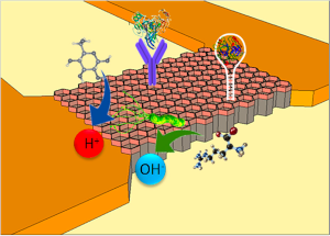
In our group, we have demonstrated the validity of multitude of nanoscopic transducers for the ultrasensitive and label-free detection of markers, in liquid(1,2) as well as in gas(3) samples, or for the microorganism monitoring in drug screening applications.(4)
In this talk, I will provide an overview of our contribution to the (bio)sensors field, including as well the use of alternative detection techniques based on memory properties(5,6), the integration with droplet microfluidics offering individual tracking of nanoliter reactors containing chemical reactions,(7) and the transfer of the transducers to flexible supports toward lower cost and light weight sensors(1,2) or for electronic skin applications.(8)
REFERENCES
1 P. Zhang, S. Yang, R. Pineda-Gómez, B. Ibarlucea, J. Ma, M. R. Lohe, T. F. Akbar, L. Baraban, G. Cuniberti and X. Feng, Small, 2019, 15, 1901265.
2 D. Karnaushenko, B. Ibarlucea, S. Lee, G. Lin, L. Baraban, S. Pregl, M. Melzer, D. Makarov, W. M. Weber, T. Mikolajick, O. G. Schmidt and G. Cuniberti, Adv. Healthc. Mater., 2015, 4, 1517–1525.
3 L. A. Panes-Ruiz, L. Riemenschneider, M. M. Al Chawa, M. Löffler, B. Rellinghaus, R. Tetzlaff, V. Bezugly, B. Ibarlucea and G. Cuniberti, Nano Res., 2022, 15, 2512–2521.
4 B. Ibarlucea, T. Rim, C. K. Baek, J. A. G. M. G. M. De Visser, L. Baraban and G. Cuniberti, Lab Chip, 2017, 17, 4283–4293.
5 B. Ibarlucea, T. F. Akbar, K. Kim, T. Rim, C.-K. C.-K. C.-K. K. Baek, A. Ascoli, R. Tetzlaff, L. Baraban, G. Cuniberti, T. Fawzul Akbar, K. Kim, T. Rim, C.-K. C.-K. C.-K. K. Baek, A. Ascoli, R. Tetzlaff, L. Baraban and G. Cuniberti, Nano Res., 2018, 11, 1057–1068.
6 B. Ibarlucea, L. Römhildt, F. Zörgiebel, S. Pregl, M. Vahdatzadeh, W. M. Weber, T. Mikolajick, J. Opitz, L. Baraban, G. Cuniberti, B. Ibarlucea, L. Römhildt, F. Zörgiebel, S. Pregl, M. Vahdatzadeh, W. M. Weber, T. Mikolajick, J. Opitz, L. Baraban and G. Cuniberti, Appl. Sci., 2018, 8, 950.
7 J. Schütt, B. Ibarlucea, R. Illing, F. Zörgiebel, S. Pregl, D. Nozaki, W. M. Weber, T. Mikolajick, L. Baraban and G. Cuniberti, Nano Lett, DOI:10.1021/acs.nanolett.6b01707.
8 B. Ibarlucea, A. Pérez Roig, D. Belyaev, L. Baraban and G. Cuniberti, Microchim. Acta, 2020, 187, 520.Speaker: Dr Bergoi Ibarlucea (Institute of Materials Science, TU Dresden, Germany) -
16:15
Atomic resolution transmission electron microscopy of radiation sensitive specimens 25m
Sub-ångström resolution has been demonstrated in transmission electron microscopy (TEM) and scanning TEM (STEM) imaging experiments thanks to the recent development of spherical and chromatic corrected equipment and/or to the development of methodologies capable to overcome the limitations related to the electron optical aberrations. Moreover, a special attention has to be paid to the eventual damage induced by the electron irradiation that can alter the structure of the specimen. Radiation damage has a dramatic sudden effect on soft matter or biologic specimens but can also affect in a subtle way the study of inorganic specimens, preventing an accurate and reliable quantification of their properties. The use of new TEM/STEM aberration corrected equipment and field emission cathodes enable to deliver a high-density of current on the specimen making radiation damage an issue of growing importance also for inorganic material and even for metals. Radiation damage is the basic handicap to atomic resolution of single particle in biology or to the development of atomic resolution methods for electron tomography. Here is discussed how in-line holography in Transmission Electron Microscopy enables the study of radiation-sensitive nanoparticles of organic and inorganic materials providing high-contrast holograms of single nanoparticles, while illuminating specimens with a density of current as low as 1 e-Å-2s s-1. This provides a powerful method for true single-particle atomic resolution imaging and opens new perspectives for the study of soft matter in biology and materials science(1). The approach is not limited to a particular class of TEM specimens, such as homogenous samples or samples specially designed for a particular TEM experiment but has better application in the study of those specimens with differences in shape, chemical composition, crystallography, and orientation, which cannot be currently addressed at atomic resolution.
1 Elvio Carlino, In-Line Holography in Transmission Electron Microscopy for the Atomic Resolution Imaging of Single Particle of Radiation-Sensitive Matter. Materials 2020, 13, 1413; doi:10.3390/ma13061413
Speaker: Dr Elvio Carlino (Institute of Crystallography, CNR, Italy) -
16:40
Controlled manipulation, lithography and sliding experiments on the nanoscale 25m
Nanotribology is a young and dynamic research field that aims to investigate friction, wear and adhesion phenomena down to the atomic scale. Since these processes occur in all natural, artificial or conceptual situations involving two surfaces (at least) in contact or in close proximity to each other, it is not surprising that, knowingly or not, many physicists, materials scientists, mechanical engineers or chemists must sooner or later confront these topics in their careers. In this talk, I will first present investigations on the friction force acting on single molecules [1], polymer chains [2] or metal clusters [3] manipulated on solid surfaces an AFM probe. If the surface itself is “manipulated”, it becomes possible to observe and quantify early stages of abrasive wear on the nanoscale, which is of utmost importance for assessing the quality of technical surfaces and possible environmental issues. Particularly instructive in this content are the cases of compliant polymers [4,5] and layered materials[6,7], where ripples, round-shaped nanoparticles, and flakes are easily generated out of the nanoscratch processes. Finally, I will also introduce first results on sliding friction in liquid environments and the influence of nanoroughness on lateral force sensing that we are currently studying in collaboration with TU Dresden.
[1] R. Pawlak, W. Ouyang, A. E. Filippov, L. Kalikhman Razvozov, S. Kawai, T. Glatzel, E. Gnecco, A. Baratoff , Q.-S. Zheng, O. Hod, M. Urbakh, E. Meyer, Single Molecule Tribology: Force Microscopy Manipulation of a Porphyrin Derivative on a Copper Surface, ACS Nano 10 (2016) 713-722
[2] J. G. Vilhena, R. Pawlak, P. D’Astolfo, X. Liu, E. Gnecco, M. Kisiel, T. Glatzel, R. Pérez, R. Häner, S. Decurtins, A. Baratoff, G. Prampolini, S. X. Liu, E. Meyer, Flexible superlubricity unveiled in sidewinding motion of individual polymeric chains, Phys. Rev. Lett. 128 (2022) 216102
[3] F. Trillitzsch, R. Guerra, A. Janas, N. Manini, F. Krok, and E. Gnecco, Directional and angular locking in the driven motion of Au islands on MoS2, Phys. Rev. B 98 (2018) 165417
[4] J. Hennig, A; Litschko, J. J. Mazo, E. Gnecco, Nucleation and detachment of polystyrene nanoparticles from plowing-induced surface wrinkling, Appl. Surf. Sci. Adv. 6 (2021) 100148
[5] J. Hennig, V. Feller, P. J. Martinez, J. J. Mazo, E. Gnecco, Locking effects in plowing-induced nanorippling of polystyrene surfaces, Appl. Surf. Sci. 594 (2022) 153467
[6] A. Özogul, F. Trillitzsch, C. Neumann, A. George, A. Turchanin, and E. Gnecco, Plowing-induced nanoexfoliation of mono- and multilayer MoS2 surfaces, Phys. Rev. Materials 4 (2020) 033603
[7] A. Özogul, E. Gnecco, and M. Z. Baykara, Nanolithography-induced exfoliation of layered materials, Appl. Surf. Sci. Adv. 6 (2021) 100146Speaker: Prof. Enrico Gnecco (Department of Solid State Physics, Jagiellonian University in Krakow, Poland)
-
15:50
-
17:05
→
17:30
Discussion and Conclusions 25m
Speakers: Dr. Cinzia Giannini (Institute of Crystallography, CNR, Italy) and Prof. Gianaurelio Cuniberti (Institute of Materials Science, TU Dresden, Germany)
-
14:30
→
15:40